Fluorescent nanoparticles and preparation method thereof

A fluorescent nanoparticle and nanoparticle technology, applied in the field of fluorescent nanoparticle and its preparation, can solve the problems of difficult application of fluorescent labeling, intolerance, poor fluorescence stability, etc., and achieve good monodispersity, uniform scale, fluorescence strong signal effect

Active Publication Date: 2018-10-16
CHONGQING UNIV
View PDF3 Cites 3 Cited by
  • Summary
  • Abstract
  • Description
  • Claims
  • Application Information

AI Technical Summary

Problems solved by technology

[0008] To sum up, the problems existing in the prior art are: affected by the environment of the solvent to be tested, fluorescent nanomaterials based on rare earth

Method used


Image

  • Fluorescent nanoparticles and preparation method thereof
  • Fluorescent nanoparticles and preparation method thereof
  • Fluorescent nanoparticles and preparation method thereof

Examples


Embodiment 1

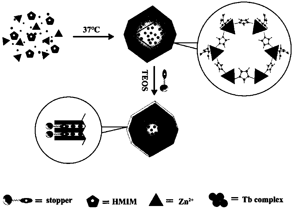
[0040] Such as figure 1 As shown, the simulation schematic diagram of the preparation method of fluorescent nanoparticles loaded with ZIF-8 rare earth complexes provided by the embodiment of the present invention, the preparation method of fluorescent nanoparticles loaded with ZIF-8 rare earth complexes provided by the present invention includes the following steps:

[0041] Step 1, pyridine two tetrazole (H 2 pytz) and triethylamine were dissolved in 1~2mL of methanol, added to a 10mL single-necked flask, mixed evenly, and then 1~4mL of terbium trifluoromethanesulfonate [Tb(OSO 2 CF 3 ) 3 ] Methanol solution in the system, stirred at room temperature for 24 hours, then vacuum-dried to remove the solvent to obtain a rare earth complex. The mass ratio of pyridine ditetrazolium, triethylamine and terbium trifluoromethanesulfonate is (43.3-129.2): (30.4-60.75): (30.3-121.2).

[0042] Step 2: Weigh 0.5 mg of the rare earth terbium complex and dissolve it in 200-300 μL of deion...

experiment example 2

[0054] The unblocked stopper nanoparticles obtained in step 2 and the fluorescent nanoparticles loaded with ZIF-8 rare earth terbium complex obtained in step 4 were respectively dispersed in PBS buffer solution with pH = 7.4, and the particle concentration was configured to be 1 mg mL -1 solution, select the excitation light wavelength as 304nm, and detect the fluorescence emission intensity of the particles at 543nm every 20 minutes to verify the stability of the fluorescence. Such as Figure 8 It can be seen from the figure that when no stopper and TEOS are added, the fluorescence intensity of the particles continues to decay over time; when the stopper and TEOS are added at a ratio of 1:2, the stability of the fluorescent nanoparticles is enhanced, and the number of It does not attenuate for hours, indicating that the particles have good tolerance to the high salt concentration and complex ion environment in the physiological system.

Embodiment 2

[0056] Step 1, pyridine two tetrazole (H 2 pytz) and triethylamine were dissolved in 1~2mL of methanol, added to a 10mL single-necked flask, mixed evenly, and then 1~4mL of terbium trifluoromethanesulfonate [Tb(OSO 2 CF 3 ) 3 ] Methanol solution in the system, stirred at room temperature for 24 hours, then vacuum-dried to remove the solvent to obtain a rare earth complex. The mass ratio of pyridine ditetrazolium, triethylamine and terbium trifluoromethanesulfonate is (43.3-129.2): (30.4-60.75): (30.3-121.2). After stirring at room temperature for 24 hours, the rare earth complex was obtained.

[0057] Step 2: Weigh 0.1 mg of the rare earth terbium complex and dissolve it in 200-300 μL of deionized water, and add it into a 10 mL single-necked flask. After uniform dispersion, add 2-methylimidazole (HMIM) with a mass of 45-54 mg and 14-16.8 mg of hexahydrate and zinc nitrate (Zn(NO 3 ) 2 ·6H 2 O). Finally, add 400-600 μL of methanol into the system and mix well, and react...


PUM


Abstract

The invention provides fluorescent nanoparticles and a preparation method thereof. The method comprises the following steps: step 1, encapsulating small molecules having fluorescent properties into pores of nanoparticles having a microporous structure; and step 2, performing a reaction on blocking molecules and the product obtained in the step 1 to obtain the fluorescent nanoparticles provided bythe invention, wherein the small molecules in the step 1 are negatively charged, and the nanoparticles are positively charged; and the blocking molecules have a molecular structure capable of blockingthe pores of the nanoparticles. According to the method provided by the invention, the obtained fluorescent nanoparticles have an average particle diameter of 100 +/- 5 nm, a specific surface area of1429 m<2>/g and a pore volume of 0.64 cm<3>/g; and the nano material has good fluorescence intensity stability, a quantum yield of 23.2%, and fluorescence lifetime of 1.9 ms, and is an ideal materialfor a biological fluorescent probe.

Description

technical field [0001] The invention belongs to the technical field of nano-biological materials, and in particular relates to a fluorescent nano-particle and a preparation method thereof. Background technique [0002] With the vigorous development of life science and technology, the research of life science has penetrated into the level of cells and single molecules of organisms. Genomics, proteomics and metabolomics methods and sequencing technologies are booming, and people have discovered more and more biomolecules such as protein markers, nucleic acids, and polypeptides that reflect physiological and pathological conditions (Nature Methods, volume 11, page 1177 ,Year 2014). The labeling, staining and detection of cells or biomolecules has become an important part of biomedical research. Among biomarkers, the most concerned labeling method is fluorescent labeling, but its detection sensitivity mainly depends on the fluorescence intensity and stability of the label. ...

Claims


Application Information

Patent Timeline
IPC IPC(8): C09K11/02C09K11/06B82Y30/00B82Y40/00
CPCB82Y30/00B82Y40/00C09K11/025C09K11/06C09K2211/182C09K2211/188
Inventor 张吉喜陈凤
Owner CHONGQING UNIV
Why Eureka
Try Eureka