Method for quantitatively analyzing tyrosine-phosphorylated proteins

A technology for quantitative analysis of tyrosine phosphorylation, applied in the field of quantitative analysis of tyrosine phosphorylated proteins, can solve the problems of inability to distinguish the occurrence of phosphorylation, and achieve the effects of saving experimental costs, convenient transformation, and clear background

Inactive Publication Date: 2018-10-23
DALIAN INST OF CHEM PHYSICS CHINESE ACAD OF SCI
View PDF3 Cites 7 Cited by
  • Summary
  • Abstract
  • Description
  • Claims
  • Application Information

AI Technical Summary

Problems solved by technology

In addition, the literature survey found that there are many relatively mature methods applied to the determination of protein concentration, such as BCA method, Coomassie brilliant blue method, etc., but there are few reported methods for the analysis of

Method used


Image

  • Method for quantitatively analyzing tyrosine-phosphorylated proteins
  • Method for quantitatively analyzing tyrosine-phosphorylated proteins
  • Method for quantitatively analyzing tyrosine-phosphorylated proteins

Examples


Embodiment 1

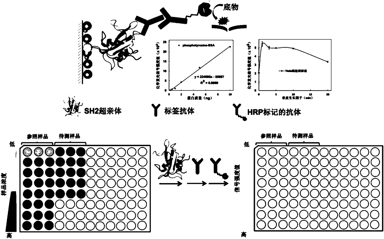
[0053] 1) Coating buffer (pH 9.6, 0.1M carbonate, freshly prepared) dilute a. tyrosine phosphorylated standard protein phosphotyrosine-BSA (SIGMA) to 0.05ng / ml, 0.25ng / ml, 0.5ng / ml ml, 1ng / ml, 2.5ng / ml, 5ng / ml, 10ng / ml, 25ng / ml, 50ng / ml, 100ng / ml, 250ng / ml, 500ng / ml, 1000ng / ml, 2500ng / ml, 5000ng / ml , b. EGF stimulated HeLa cell lysate for 1 minute to 250ng / ml, 500ng / ml, 1000ng / ml, 2500ng / ml, 5000ng / ml, c. 2000ng / ml alkaline phosphatase treatment for 0 minutes, 3 minutes, 5 minutes Minutes, 10 minutes, 20 minutes, 30 minutes, 60 minutes of HeLa cell lysate stimulated with epidermal growth factor for 20 minutes, the above samples were added to a 96-well plate at 100 μL / well, and each sample was added to three wells in parallel, at 37 degrees Celsius Incubate for 2 hours;

[0054] 2) Remove the solution in step 1), wash with TBST (20mM Tris, 150mM NaCl, pH 7.4, 0.05% (v / v) Tween-20) three times, 200μL / time, 3min / time, and finally wash in a dust-free pat dry on paper;

[0055] ...

Embodiment 2

[0069]1) Coating buffer (pH 9.6, 0.1M carbonate, freshly prepared) serially dilute tyrosine phosphorylated standard protein phosphotyrosine-BSA (SIGMA) to 0.1ng / ml, 0.25ng / ml, 0.5ng / ml , 1ng / ml, 2.5ng / ml, 5ng / ml, 10ng / ml, 25ng / ml, 50ng / ml and 50ng / ml, 100ng / ml, 250ng / ml, 500ng / ml, 1000ng / ml, 2500ng / ml, Add 5000ng / ml, 100μL / well into a 96-well plate, add each sample to three wells in parallel, and incubate at 37°C for 2 hours;

[0070] 2) Remove the solution in step 1), wash three times with TBST (20mM Tris-HCl, 150mM NaCl, pH7.4, 0.05% (v / v) Tween-20), 200μL / time, 3min / time, and finally Pat dry on a dust-free paper;

[0071] 3) Add 200 μL of blocking solution (TBST containing 5 wt.% skimmed milk powder) to the 96-well plate after being patted dry in step 2), and react at 37 degrees Celsius for 1 hour;

[0072] 4) After the reaction, wash the 96-well plate according to step 2);

[0073] 5) Add 100 μL of mouse tyrosine phosphorylated antibody P-Tyr-100 (1:1000) to the 96-well...

Embodiment 3

[0081] First, biotinylate the SH2 superphile, the specific operation is

[0082] 1) Take 2ml of 2mg / ml SH2 superphile and biotinylation reagent such as biotin-NHS molar ratio 1:5, shake and react in the dark, 4°C overnight; add 100μL NH 4 Cl terminated the reaction for 30min;

[0083] 2) Ultrafilter the solution after the reaction in step 2) with a 10k ultrafiltration tube, wash with PBS three times, then invert the ultrafiltration tube to collect the liquid.

[0084] 3) The concentration of the solution (SH2-biotin) after the SH2 superphile and biotin cross-linked obtained in step 3) by BCA method;

[0085] 4) The SH2-biotin solution obtained in step 3) of the HABA method measures the average number of labeled biotin per SH2;

[0086] 5) ELISA method to evaluate the best ratio;

[0087] 6) Aliquot the SH2-biotin solution obtained in step 3) and freeze at -80°C.

[0088] Secondly, use biotinylated SH2 superphile as the primary antibody to quantitatively analyze the level o...


PUM

PropertyMeasurementUnit
Concentrationaaaaaaaaaa

Abstract

The invention relates to a method for quantitatively analyzing tyrosine-phosphorylated proteins. The method is characterized in that the dynamic change curve of the tyrosine phosphorylation level of asample to be tested is obtained based on an ELISA technology with a modified SH2 over parent as a first antibody, a standard curve is obtained with tyrosine-phosphorylated standard proteins as a reference sample in the detection process, and the quantitative analysis of the tyrosine phosphorylation level of the sample to be tested is realized with the standard curve as a reference. The method hasthe advantages of low cost, no sample pre-enrichment, good specificity and low detection limit. The ELISA technology is carried out in a well plate, and an apparatus is simple. The SH2 over parent adopted in the invention can be used to obtain a lot of proteins with a stable structure through bacterium expression and purification, so the experimental cost is greatly saved, the background is clear, and reconstruction is easy. The method has the same high affinity and high specificity to the tyrosine-phosphorylated proteins as antibody, has a broad spectrum, and can successfully realize the quantitative analysis of the tyrosine phosphorylation level of multiple complex samples.

Description

technical field [0001] The invention belongs to the technical field of tyrosine phosphorylation proteomics in the direction of proteomics research, and specifically relates to a quantitative analysis method for tyrosine phosphorylation proteins. Background technique [0002] The tyrosine phosphorylation signal transduction system is involved in many important cell biological functions, such as cell differentiation, proliferation, migration and apoptosis (literature: Hunter, T., Cell 2000, 100, 113–127.), when it occurs When it is abnormal, it is often accompanied by the occurrence of diseases, especially cancer (document: Rix, U.; Superti-Furga, G., Nat. Chem. Biol. 2009, 5, 616-624.). Therefore, the research on tyrosine phosphorylation signaling pathway is a focus in the field of biomedicine. Up to now, among the 26 kinase inhibitors approved by the US Food and Drug Administration (FDA) for cancer targeted therapy, 22 One (84%) targets phosphorylated tyrosine kinases (arti...

Claims


Application Information

Patent Timeline
IPC IPC(8): G01N33/535G01N33/543
CPCG01N33/535G01N33/54306
Inventor 叶明亮李亚楠
Owner DALIAN INST OF CHEM PHYSICS CHINESE ACAD OF SCI
Why Eureka
Try Eureka