Application of compound as blocking reagent for glycosyl on antibody

A technology for blocking reagents and compounds, applied in the field of protein glycosyl detection, can solve problems such as interference in detection results, and achieve the effects of accurate detection and analysis, good activity, and shortening the reaction process.

Inactive Publication Date: 2018-11-13
嵊州宝康医疗科技有限公司
View PDF4 Cites 2 Cited by
  • Summary
  • Abstract
  • Description
  • Claims
  • Application Information

AI Technical Summary

Problems solved by technology

However, since the antibody itself has glycosyl groups, it will greatly interfere with the detection results

Method used


Image

  • Application of compound as blocking reagent for glycosyl on antibody
  • Application of compound as blocking reagent for glycosyl on antibody
  • Application of compound as blocking reagent for glycosyl on antibody

Examples


Embodiment 1

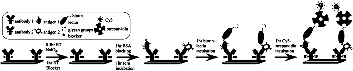
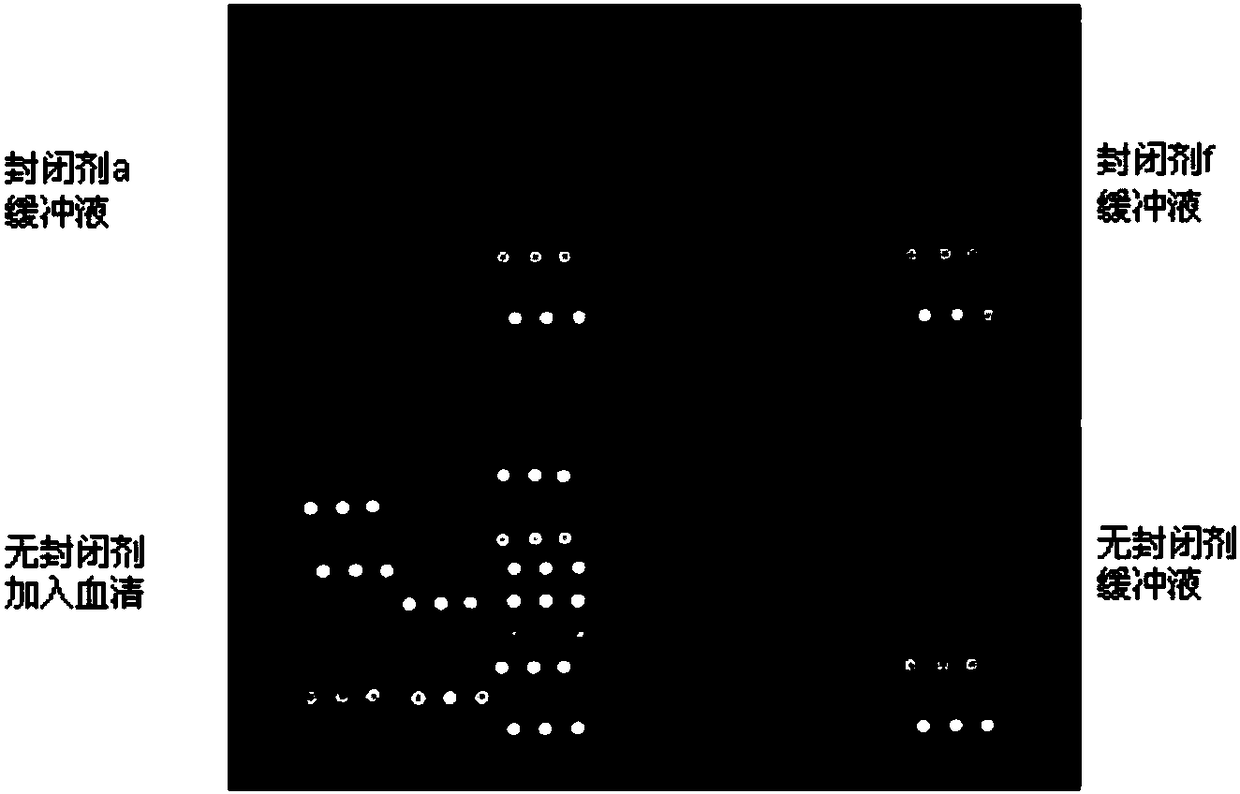
[0067] Coat the antibody onto the surface of the antibody chip, add 150 mmol of sodium periodate, add 20 mmol of pH7.0 sodium acetate buffer, and react at 4°C for 2 hours; add blocking reagent a, and use the same at room temperature The buffer was incubated for 1 hour; the surface of the antibody chip was blocked with calf serum for 1 hour; at room temperature, biotin-labeled lectin AAL was added and incubated for 1 hour; then Cy3-labeled streptavidin was added and incubated for 1 hour Carry out microarray scanning detection to the prepared antibody chip, detect the amount of the combined lectin with Cy3 fluorescent dye, the detection result sees image 3 The upper left subarray in .

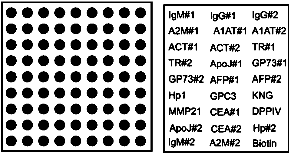
[0068] figure 2 For the array map of antibodies, each antibody was replicated 3 times to generate 3 dots with a diameter of 130 μm. Each point is represented by (column, row), such as the first antibody in the first row is represented as IgM#1(1,1)(2,1)(3,1).

[0069] image 3 BiotinBSA(7,9...

Embodiment 2

[0073] Coat the antibody onto the surface of the antibody chip, add 150 millimolar sodium periodate in 20 millimolar pH7.0 sodium acetate buffer and react at 4°C for 2 hours; add blocking reagent f, and use the same buffer at room temperature The solution was incubated for 1 hour; the surface of the antibody chip was blocked with calf serum for 1 hour; at room temperature, biotin-labeled phytohemagglutinin AAL was added and incubated for 1 hour; then Cy3-labeled streptavidin was added and incubated for 1 hour; Perform microarray scanning detection on the prepared antibody chip, and use Cy3 fluorescent dye to detect the amount of bound lectin. The detection results are shown in image 3 The upper right subarray of , figure 2 An array map of antibodies.

[0074] Such as image 3 As shown in the upper right subarray, when the glycosyl on the antibody was blocked with the blocking agent f, most of the antibodies incubated with phosphate buffer did not develop color, indicating ...

Embodiment 3

[0077] Coat the antibody to the surface of the antibody chip; block the sugar group on the antibody with blocking agent a or blocking agent f, and block the surface of the antibody chip with calf serum for 1 hour; add serum or buffer, at room temperature, add biotin Labeled recombinant lectin AAL (for detection of trehalose), wild plant lectin AAL (for detection of trehalose), phytohemagglutinin RCA120 (for detection of glucose amide), or phytohemagglutinin SNA (for detection of sialic acid), incubated for 1 hour; Add Cy3-labeled streptavidin and incubate for 1 hour; perform microarray scanning detection on the prepared antibody chip, and detect the amount of bound lectin with Cy3 fluorescent dye.

[0078] Such as Figure 4 As shown, after blocking the sugar group on the antibody with blocking agent f, when the recombinant phytohemagglutinin AAL (AleuriaAurantia Lectin orange reticulocyte agglutinin) is used to detect, the positive control wells develop color, and the negative...


PUM


Abstract

The invention relates to the detection of protein glycosyl, in particular to an application of a compound as a blocking reagent for glycosyl on an antibody. To solve the problem of the interference ofthe glycosyl of a current antibody on detection, a compound used as a blocking reagent for the glycosyl on the antibody is provided, and the compound is selected from one of alpha-semicarbazide, acetylhydrazine, sulfur acetohydrazide, thiosemicarbazide, asparagine hydrazide, tyrosine hydrazine, N (benzyloxycarbonyl)-L-valyl-L-tyrosine hydrazide, or N (benzyloxycarbonyl) glycinic hydrazide. The compound used as the blocking reagent for the glycosyl on the antibody has the advantages of being capable of conducting petreatment of antibodies, avoiding binding of plant lectins to antibodies, guaranteeing accurate quantification of the glycosyl on captured proteins, being simple in the process of blocking the antibodies with one step reaction needed, shortening the reaction process of existingblocked antibodies, and significantly reducing costs.

Description

technical field [0001] The present invention relates to the detection of protein glycosyl groups, in particular to the application of a compound as a blocking reagent for glycosyl groups on antibodies. Background technique [0002] Early diagnosis of disease using molecular markers can significantly reduce the death caused by the disease. Glycosylation of proteins is the most common post-translational modification of proteins. These modifications play an important role in the physical, chemical and biological properties of proteins; at the same time, they play a very important role in various biological processes, including development, cell differentiation, fertilization, cellular immune recognition, host-pathogen interaction, and intercellular signal transduction role. Because the sugar group is particularly sensitive to the environment. Therefore, the detection of abnormal glycosyl groups can be used as a specific biomarker for diagnosing diseases, and can be used for ...

Claims


Application Information

Patent Timeline
Patent Type & Authority Applications(China)
IPC IPC(8): C07C281/06C07C243/28C07C327/56C07C337/06C07C243/34C07C271/22C07K5/062G01N33/68
CPCC07C243/28C07C243/34C07C271/22C07C281/06C07C327/56C07C337/06C07K5/06078G01N33/68
Inventor 陈松明
Owner 嵊州宝康医疗科技有限公司
Why Eureka
Try Eureka