Extracellular vesicle and inclusion extraction method and device based on electrostatic adsorption

An extraction device and electrostatic adsorption technology, applied in the field of extracellular vesicle separation and extraction, can solve the problems of low yield, cumbersome operation, and low sample purity

Inactive Publication Date: 2018-11-20
北京恩泽康泰生物科技有限公司
View PDF4 Cites 8 Cited by
  • Summary
  • Abstract
  • Description
  • Claims
  • Application Information

AI Technical Summary

Problems solved by technology

[0005] Specifically, the present invention addresses the common problems of cumbersome operation, low yield, and low samp

Method used


Image

  • Extracellular vesicle and inclusion extraction method and device based on electrostatic adsorption
  • Extracellular vesicle and inclusion extraction method and device based on electrostatic adsorption
  • Extracellular vesicle and inclusion extraction method and device based on electrostatic adsorption

Examples


Embodiment 1

[0063] Example 1 Adsorption of anion exchange resin particles to extracellular vesicles

[0064] 1. Preparation of reagents and consumables

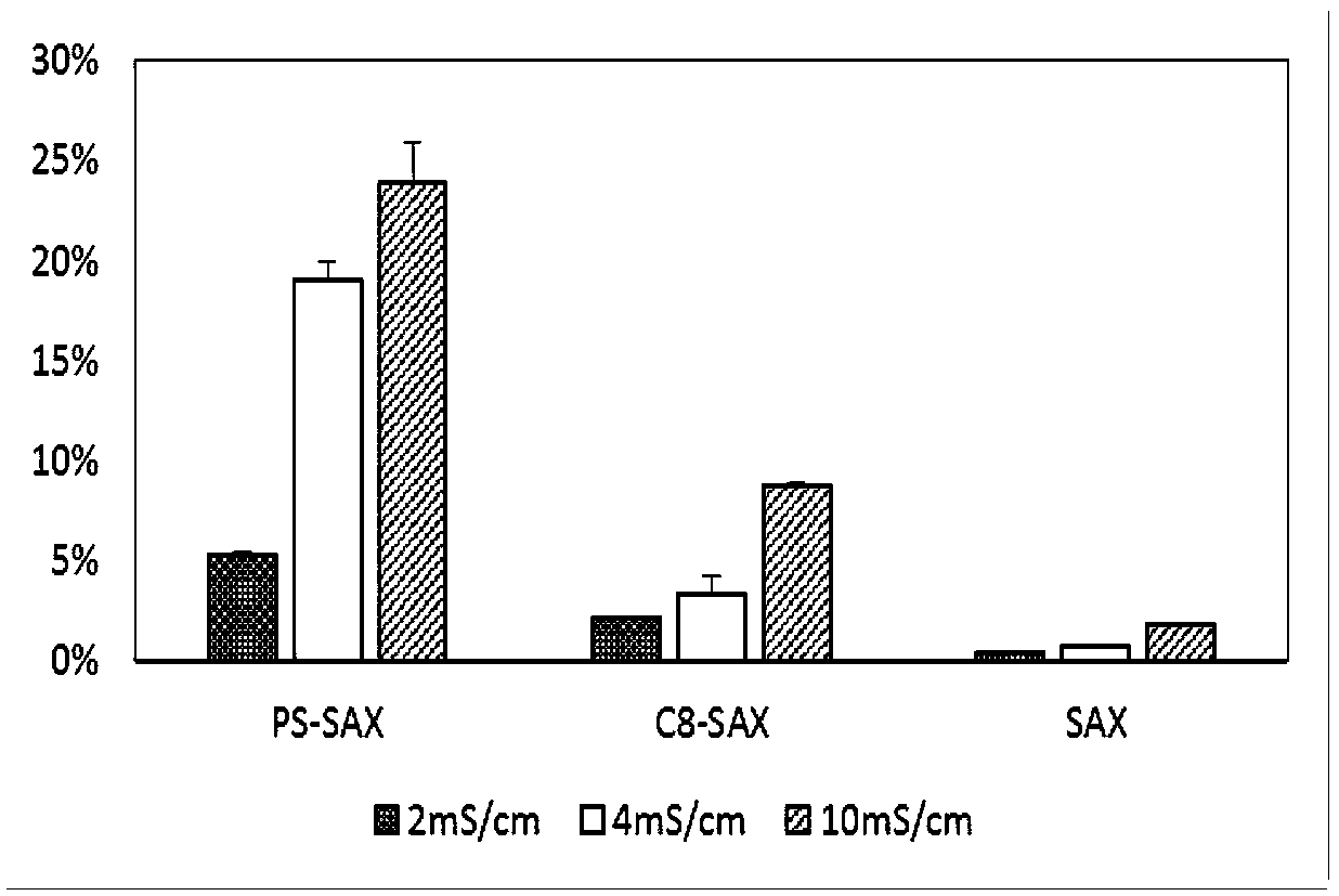
[0065] The anion exchange resin particles used include polystyrene particles with quaternary ammonium functional groups (PS-SAX), silica gel particles with C8 and quaternary ammonium functional groups (C8-SAX) and silica gel particles with quaternary ammonium functional groups (SAX), particle size Between 30-800μm. Cell membrane green fluorescent probe DIO. Commercial human serum. Dilute the PBS solution at pH 7.4 with DI water to prepare buffer solutions with conductivity of 2mS / cm, 4mS / cm and 10mS / cm respectively.

[0066] 2. Preparation of green fluorescently labeled extracellular vesicles (fEVs)

[0067] Thawed commercial human plasma was centrifuged at 1300g for 15 minutes to remove cytoplasmic debris. Then transfer the supernatant to a new centrifuge tube, centrifuge at 3000g for 15 minutes, mix the supernatant with PBS at a v...

Embodiment 2

[0071] Example 2 Adsorption effect of different device forms on fEVs

[0072] 1. Preparation of reagents and consumables

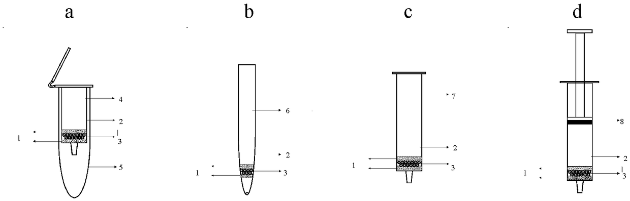
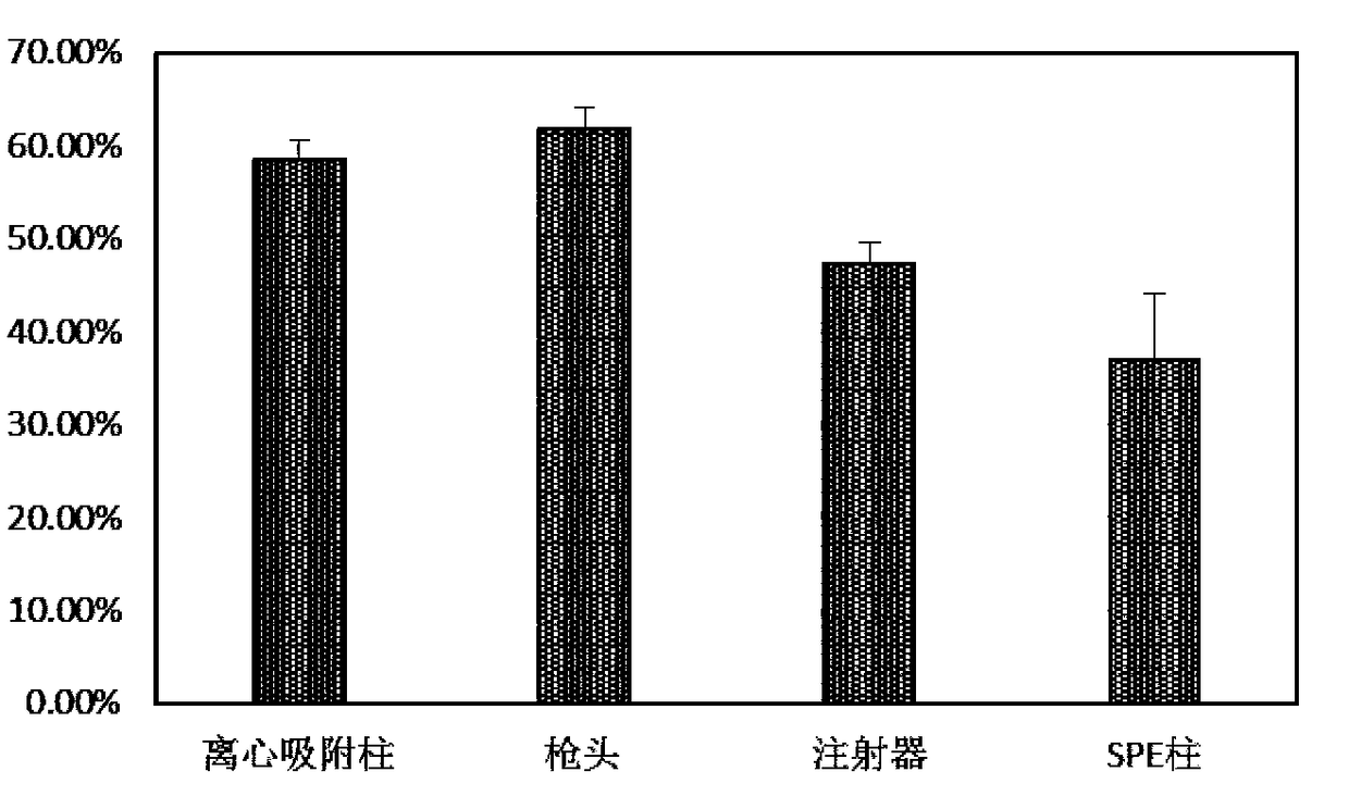
[0073] The anion exchange resin particles used are silica gel particles (SAX) with quaternary ammonium functional groups, and the particle size is between 30-800 μm. Green fluorescently labeled extracellular vesicles (fEVs) were prepared in the same way as in Experimental Example 1. The centrifugal adsorption column used is a centrifugal empty column with an outer tube volume of 15 mL. Use a pipette with a standard 1mL tip. The syringe used is a 1mL disposable syringe. The solid phase extraction column (SPE column) used was an empty SPE column (12 mL) with an inner diameter of 13 mm. Unless otherwise specified, the sieves used are all hydrophobic sieves with a pore size of 10 μm. The specific assembly method is as figure 1 shown.

[0074] 2. Adsorption of fEVs by anion exchange resin particles with different assembly forms

[0075]Add fEVs to PBS b...

Embodiment 3

[0076] The influence of embodiment 3 conductivity on EVs trapping effect

[0077] 1. Preparation of reagents and consumables

[0078] The centrifugal adsorption column used is a centrifugal empty column with an outer tube volume of 15 mL, and the selected sieve plate is a hydrophobic sieve plate with a pore size of 10 μm. The used anion exchange resin particles are silica gel particles (SAX) with quaternary ammonium functional groups, and the particle diameter is 30-800 μm. The biological sample used is the supernatant of 22rv1 cells cultured in exosome-free fetal bovine serum medium, which is filtered through a 0.45 μm filter membrane before use. Use DI water and NaCl to adjust the conductivity of the cell supernatant between 4-80mS / cm. The kit used for EVs nucleic acid extraction was miRNeasy Mini Kit (Qiagen). The obtained RNA was analyzed with Agilent RNA 6000 Pico Kit.

[0079] 2. Capture EVs in cell supernatant and extract RNA

[0080] Such as figure 1 Assemble the...


PUM

PropertyMeasurementUnit
Particle sizeaaaaaaaaaa
Conductivityaaaaaaaaaa

Abstract

The invention provides an extracellular vesicle and inclusion extraction method and device based on electrostatic adsorption. After being pretreated, a biological sample is added into an extraction device, in which an anion exchange resin particle is arranged; effluent is discharged from a port in the bottom of the extraction device; the extraction device is cleaned with a cleaning buffer solutionand can be eluted with an eluting buffer solution; eluted extracellular vesicles are collected, or extracellular vesicles captured in the resin are directly split to extract inclusions. The surface of the anion exchange resin particle is modified by at least one of the following functional group: quaternary ammonium, tertiary amine, secondary amine and primary amine. By full contact between the biologically sample and the anion exchange resin particle, the extracellular vesicles with negative charges on the surface are adsorbed onto the surface of the particle under the electrostatic action,so as to realize quick and efficient separation. The extracellular vesicles are separated by utilizing electrostatic adsorption, and over 50 percent of extracellular vesicles can be recycled from various biological samples within 30 minutes.

Description

technical field [0001] The invention relates to the separation and extraction technology of extracellular vesicles, in particular, a method and device for extracting extracellular vesicles and their contents based on electrostatic adsorption. Background technique [0002] Cells can secrete a variety of vesicles with a phospholipid bilayer structure, including endosome-derived exosomes (Exosome, particle size 50-100nm) and plasma membrane-derived microvesicles (Microvesicle, particle size 20-1000nm) . Extracellular vesicles have been shown to play roles in blood coagulation, intercellular signaling, and waste management. 1 At the same time, extracellular vesicles have broad application prospects in the diagnosis and treatment of diseases because they are rich in nucleic acids, proteins, lipids and other biomolecules. [0003] At present, the extraction and separation methods of extracellular vesicles mainly include ultracentrifugation, size exclusion chromatography, immunoc...

Claims


Application Information

Patent Timeline
IPC IPC(8): C12N5/00C12M1/00
CPCC12M47/04C12N5/0602
Inventor 孔关义赵立波陈苏红杨玉清李志
Owner 北京恩泽康泰生物科技有限公司
Why Eureka
Try Eureka