Kit and method for preparing exosomes in serum or plasma
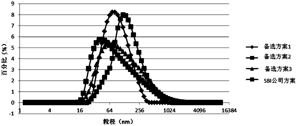
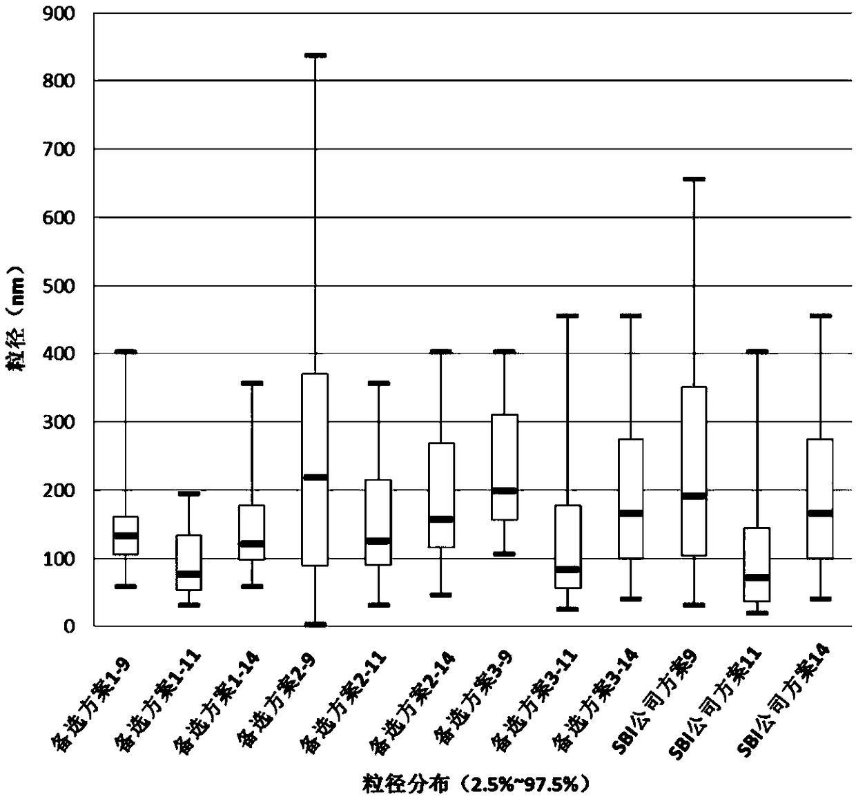
A technology of exosomes and kits, applied in the field of biomedicine, can solve the problems of large particle range, low yield, and low purity, and achieve the effects of concentrated particle size distribution, reduced interference, and guaranteed activity
- Summary
- Abstract
- Description
- Claims
- Application Information
AI Technical Summary
Problems solved by technology
Method used
Image
Examples
preparation example Construction
[0069] In addition, the present invention also provides a method for preparing exosomes in serum or plasma, comprising the following steps:
[0070] Add any of the above-mentioned exosome extraction reagents to the serum or plasma sample, mix well, let stand, and centrifuge to remove the supernatant to obtain exosome precipitation; the amount of exosome extraction reagent is 1 / 2 of the volume of the sample to be extracted. 1 / 4 times - 1 times, preferably 1 / 4 times - 1 / 2 times; the condition of standing still is preferably 4 ℃ for 10-60 minutes, more preferably 20-40 minutes, when sufficient settlement is ensured At the same time, the activity of exosomes is guaranteed; the centrifugation condition is preferably 2000g-15000g for 10 minutes, more preferably 8000g-12000g for 10 minutes.
[0071] Add the above-mentioned exosome lysis reagent to the exosome precipitation to obtain an exosome lysis solution; the exosome lysis reagent is preferably PBS buffer solution.
[0072] Add ...
Embodiment 1
[0087] Extraction of exosomes from serum
[0088] The composition of the reagents used:
[0089] Extraction reagent P1 (dextran sulfate 13,000, 5%, glucose, 2.5%)
[0090] Dissolving reagent S1 (PBS solution)
[0091] Purified Microspheres B1 (Protein G and Butyl-Sepharose 4B, 20:1)
[0092] 1. Centrifuge at 3,000g 4°C for 15 minutes to remove residual cells and debris in the serum.
[0093] 2. Add 25 μl P1 to 100 μl serum for each sample, mix by inversion, let stand at 4°C for 30 minutes, and centrifuge at 10,000 g for 10 minutes.
[0094] 3. Discard the supernatant, collect the precipitate, centrifuge again at 10,000g for 5 minutes, and discard the remaining supernatant.
[0095] 4. Add the S1 solution twice the initial volume, and gently pipette to dissolve to obtain the exosome solution.
[0096] 5. Take out purified microspheres B1 equal in volume to the initial plasma volume, centrifuge at 4,000g for 3 minutes, discard the supernatant, add 1ml of S1 solution and mix...
Embodiment 2
[0100] Extraction of exosomes from plasma
[0101] The composition of the reagents used:
[0102] Extraction Reagent P1 (PEG 30,000, 5%, Dextran Sulfate 15,000, 5%, Glucose, 6%)
[0103]Dissolving reagent S1 (PBS solution)
[0104] Purification Microspheres B1 (Protein G and Butyl-Sepharose 4B, 1:1)
[0105] Proteinase K (20%)
[0106] 1. Centrifuge at 3,000g 4°C for 15 minutes to remove residual cells and debris in the serum.
[0107] 2. Transfer the supernatant to a new EP tube, add 0.5 times the volume of S1 solution, invert or vortex to mix.
[0108] 3. Add 0.05 times the volume of Proteinase K, mix the sample, incubate at 37°C for 10 minutes, and then pre-cool to 4°C.
[0109] 4. Add 0.2 times the volume P1 of the existing total volume (volume of plasma + volume of S1) to the sample, mix well and place at 4°C for 30 minutes.
[0110] 5. Centrifuge at 10,000 g at 4°C for 10 minutes.
[0111] 6. Collect the precipitate, discard the supernatant, centrifuge again at 10...
PUM
| Property | Measurement | Unit |
|---|---|---|
| molecular weight | aaaaa | aaaaa |
| molecular weight | aaaaa | aaaaa |
| molecular weight | aaaaa | aaaaa |
Abstract
Description
Claims
Application Information
 - R&D
- Intellectual Property
- Life Sciences
- Materials
- Tech Scout
- Unparalleled Data Quality
- Higher Quality Content
- 60% Fewer Hallucinations
Browse by: Latest US Patents, China's latest patents, Technical Efficacy Thesaurus, Application Domain, Technology Topic, Popular Technical Reports.
© 2025 PatSnap. All rights reserved.Legal|Privacy policy|Modern Slavery Act Transparency Statement|Sitemap|About US| Contact US: help@patsnap.com


