Miniaturized multi-angle three-dimensional super-resolution light sheet fluorescence microscope

A fluorescence microscope and super-resolution technology, applied in microscopes, optics, optical components, etc., can solve the problems of unsatisfactory spatial resolution, bulky, and expensive, and achieve compact structure, optimized design, and low cost.

Active Publication Date: 2018-12-14
HUAZHONG UNIV OF SCI & TECH
View PDF2 Cites 14 Cited by
  • Summary
  • Abstract
  • Description
  • Claims
  • Application Information

AI Technical Summary

Problems solved by technology

For developmental biology, neurobiology, pathology and other fields that require frequent observation of embryos, tissues, organs and other large samples with a scale of several millimeters, the spatial resolution is still difficult to meet the requirements
[0004] At the same time, the existing commercial light-sheet fluorescence microscope is expensive, costing millions of dollars, and the operation is cumbersome and bulky

Method used


Image

  • Miniaturized multi-angle three-dimensional super-resolution light sheet fluorescence microscope
  • Miniaturized multi-angle three-dimensional super-resolution light sheet fluorescence microscope
  • Miniaturized multi-angle three-dimensional super-resolution light sheet fluorescence microscope

Examples


Embodiment 1

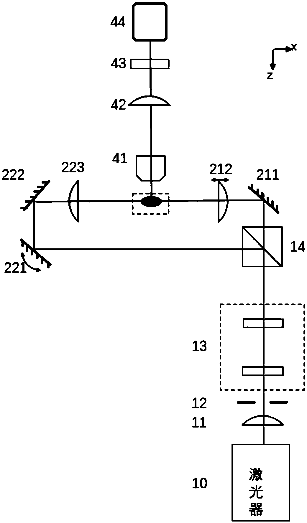
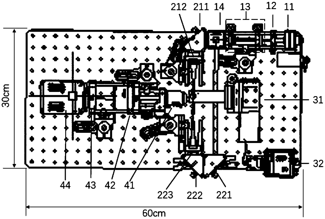
[0053] The miniaturized multi-angle three-dimensional super-resolution light-sheet fluorescence microscope provided in this embodiment makes the device more compact through the improvement of the optical path and the selection of devices. This miniaturized device realizes multi-angle three-dimensional super-resolution light sheet microscopy imaging on an optical platform of only 30*60cm.

[0054] The overall structure of the miniaturized multi-angle three-dimensional super-resolution light sheet microscope device is as follows: image 3 shown. 11 is a fiber collimator with a focal length of 15mm; 12 is an adjustable slit diaphragm with an effective adjustment range of 0-8mm; 13 is a beam expander shaping module, and the focal lengths of two cylindrical mirrors are 9.7mm and 25.4mm; 14 211 is the first reflector, 212 is a cylindrical mirror with a focal length of 75mm; 221 is the second reflector and 222 is the third reflector, and 223 is a cylindrical mirror with a focal leng...

Embodiment 2

[0059] In this embodiment, the device is mainly used to image and detect microemulsion droplets. Usually the size of microemulsion droplets is between tens to hundreds of microns, and the spatial resolution is not very high, so super-resolution is not required; due to the scattering of droplets, it is difficult to scan through the entire sample with a single illumination (the place where the droplets are placed) The maximum diameter of the centrifuge tube is about 3mm), so multi-angle fusion is required. The device can realize rapid high-throughput imaging detection of a large number of microemulsion droplets.

[0060] Image 6 It is the result of multi-channel three-dimensional imaging of liquid droplets marked by FAM / HEX staining using the device. FAM is excited by blue light, and HEX is excited by green light. The excitation wavelengths used were 488nm and 532nm respectively, the imaging exposure time was set to 100ms, and the scanning step was set to 10μm. Three hundred...

Embodiment 3

[0064] This example is the imaging of transgenic mouse cranial nerves labeled with green fluorescent protein. Thy-1 genotype green fluorescent protein is expressed in mouse brain neurons and nerve fibers. Due to the opacity of mouse brain tissue, it is difficult to observe with an optical microscope, so it needs to be cleared first, such as Figure 8 a, b. Even after clearing, deep image degradation is still severe due to scattering and absorption by tissue. Figure 8 In c, the imaging result of a conventional light sheet microscope is shown. The original xy two-dimensional image is shown on the left, and the xz reconstruction surface is shown on the right. It can be seen that after clearing, even if it is imaged with an advanced light sheet microscope, the degradation effect of the deep tissue is still obvious, and because the axial resolution is larger than the lateral resolution, the axial direction is elongated. The effect is reflected on the xz surface. Figure 8 d an...


PUM


Abstract

The invention discloses a miniaturized multi-angle three-dimensional super-resolution light sheet fluorescence microscope. The microscope comprises a light source module, a light sheet generation module, a sample control module and an image acquisition module; the light source module is used for forming a beam of collimated elliptical light; the light sheet generation module is used for producinga light sheet according to the collimated elliptical light; the sample control module is used for controlling a sample to move to be scanned by the light sheet when the light sheet is irradiated on the sample; and the image acquisition module is used for synchronously acquiring fluorescence light for exciting the sample to form an image sequence. The microscope has relatively high cost performance; relatively few elements are adopted; a hardware device of the microscope is small and exquisite compared with a large-scale Bezier light sheet microscope, a commercial light sheet microscope and thelike; and the microscope is simple in operation and has great practical values.

Description

technical field [0001] The invention belongs to the field of microscopic imaging, and more specifically relates to a miniaturized multi-angle three-dimensional super-resolution light-sheet fluorescence microscope. Background technique [0002] Light-sheet fluorescence microscopy is a new type of microscopic imaging technology emerging in this century. Unlike traditional epi-fluorescence microscopy, the illumination and acquisition of light-sheet fluorescence microscopy are independent of each other, that is, the illumination source and detection acquisition are perpendicular to each other. A sheet-shaped light source is used to excite the fluorescence of the sample from the side, and the excited fluorescence image of the illuminated layer is collected by using a wide field, and the two-dimensional image sequence is obtained by scanning to reconstruct the three-dimensional. Compared with ordinary wide-field fluorescence microscope and laser scanning confocal microscope, ligh...

Claims


Application Information

Patent Timeline
Patent Type & Authority Applications(China)
IPC IPC(8): G02B21/00G02B21/26
CPCG02B21/0032G02B21/0036G02B21/0076G02B21/26
Inventor 费鹏聂俊朱兰馨
Owner HUAZHONG UNIV OF SCI & TECH
Why Eureka
Try Eureka