Application of self-assembling oligopeptide to rapid repair of skin tissue trauma
A technology of self-assembling short peptides and tissue trauma, which is applied in the field of nanobiological regenerative medicine, can solve the problems of poor curative effect and slow repair of skin tissue damage, and achieve the effect of less inflammation, faster wound healing and better repair quality
- Summary
- Abstract
- Description
- Claims
- Application Information
AI Technical Summary
Problems solved by technology
Method used
Image
Examples
Embodiment 1
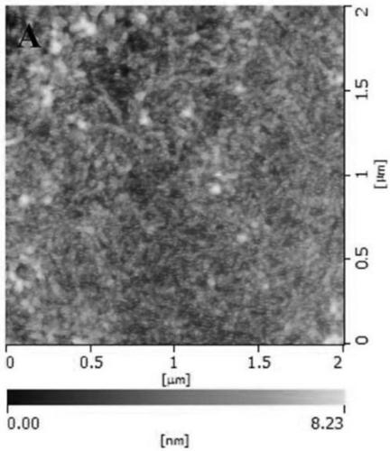
[0038]Example 1: Atomic force microscopy (AFM) of self-assembled short peptide Sciobio-II self-assembled for 24h
[0039] The self-assembled short peptide after self-assembly at 37°C for 24 hours was shown in the atomic force microscope. After 24 hours of assembly, the short peptide formed a dense nanorod-like fiber structure, and the rod-like nanofibers were assembled into a dense nanofiber network scaffold. , to provide support and adhesion for the three-dimensional culture of cells ( figure 1 ).
Embodiment 2
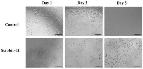
[0040] Embodiment 2: The cultivation of HFF-1 cells in the three-dimensional environment constructed by Sciobio-II
[0041] Human skin fibroblasts (HFF-1) were routinely cultured in a two-dimensional environment, high-glucose DMEM medium with 15% FBS and 1% double antibody, CO 2 Culture in a cell culture incubator (37°C, 5%). The three-dimensional culture steps are as follows: (1) Cells are adhered to grow to about 80% for three-dimensional culture; (2) Wash the cells twice with PBS before digestion with trypsin (0.25%); (3) Centrifuge at 1000rpm and count; (4) Add short Peptide and other solutions, mix well to form a three-dimensional suspension cell solution. Place the 96-well plate in a constant temperature incubator (37°C, 5% CO 2 ) for cultivation, observation and analysis. In a two-dimensional environment, HFF-1 is in a state of adherent growth, showing a long fusiform shape. When cultured in the three-dimensional environment constructed by Sciobio-II, the cells are ...
Embodiment 3
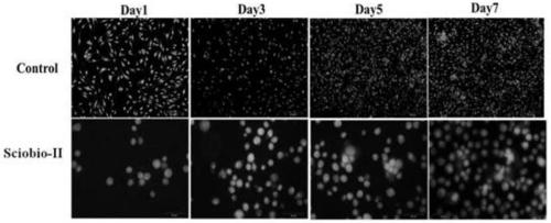
[0042] Example 3: Acridine orange / ethidium bromide staining of HFF-1 cells in the three-dimensional environment constructed by Sciobio-II
[0043] The steps are as follows: (1) HFF-1 cells were cultured in the two-dimensional environment and the three-dimensional environment constructed by Sciobio-II. About 2000 cells were seeded per well. The well plate was placed at 37°C, 5% CO 2 cultured in a constant temperature incubator. 24h after plating was recorded as the first day, and AO / EB staining was carried out on the 1st, 3rd, 5th and 7th day; (2) AO dyeing solution and EB dyeing solution were mixed evenly in the dark to make AO / EB dyeing solution; ( 3) Take the well plate and discard the culture medium. Wash with PBS. Fix with 20 μl / well of 4% paraformaldehyde for 10-15 minutes. Add 15 μl / well of AO / EB staining solution in the dark, and stain for 10 minutes. Use PBS to wash away the background. Observed and photographed under a fluorescent inverted microscope.
[0044]...
PUM
 Abstract
Description
Claims
Application Information
 - R&D
- Intellectual Property
- Life Sciences
- Materials
- Tech Scout
- Unparalleled Data Quality
- Higher Quality Content
- 60% Fewer Hallucinations
Browse by: Latest US Patents, China's latest patents, Technical Efficacy Thesaurus, Application Domain, Technology Topic, Popular Technical Reports.
© 2025 PatSnap. All rights reserved.Legal|Privacy policy|Modern Slavery Act Transparency Statement|Sitemap|About US| Contact US: help@patsnap.com


