A method and device for displaying hard tissue pathological slices

A technology for pathological slices and hard tissues, applied in the field of pathological tissue slice preparation, which can solve problems such as poor patch effect, tissue rupture, and incomplete slices, and achieve the effects of short processing time, easy operation, and uniform pressure

Active Publication Date: 2021-01-29
XIANGYA HOSPITAL CENT SOUTH UNIV
View PDF4 Cites 0 Cited by
  • Summary
  • Abstract
  • Description
  • Claims
  • Application Information

AI Technical Summary

Problems solved by technology

This method solves technical problems such as poor patch effect, incomplete slice, slice shrinkage and tissue rupture caused by sticking slices in hard tissue slices, and the invention is simple and convenient to operate, with obvious slice spreading effect and good repeatability

Method used


Image

  • A method and device for displaying hard tissue pathological slices
  • A method and device for displaying hard tissue pathological slices
  • A method and device for displaying hard tissue pathological slices

Examples


Embodiment 1

[0025] 1) Obtaining materials and fixing: take mouse knee joints, carefully remove the surrounding attachment tissues, place them in an appropriate amount of 4% paraformaldehyde and fix them for 24 hours (the femur and tibia form an angle of 90-120°);

[0026] 2) Flushing: Rinse the fixed joint tissue in running water for 10 minutes, soak in PBS for 10 minutes, wash off the fixative, and use absorbent paper to absorb excess liquid around the knee joint tissue;

[0027] 3) Dehydration and transparency: the knee joint tissues after flushing were placed in 30%, 50%, 70%, 80%, 95% Ⅰ, 95% Ⅱ, 100% Ⅰ, 100% Ⅱ alcohol for gradient dehydration. Drain alcohol for 24 hours; place in xylene I and xylene II in turn for tissue transparency, each xylene for 12 hours;

[0028] 4) Permeation: The transparent knee joint tissue was placed in permeation solution I (methyl methacrylate / dibutyl phthalate volume ratio: 2:1), permeation solution II (methyl methacrylate / ortho The volume ratio of dibut...

Embodiment 2

[0037] 1) Deplasticization: Place the hard tissue slides removed from vaseline in 2-methoxyethyl acetate Ⅰ, 2-methoxyethyl acetate Ⅱ, 2-methoxyethyl ethyl acetate in sequence. Tissue demodeling in Ester III, 20 minutes per pass with 2-methoxyethyl acetate;

[0038] 2) Rehydration: place the deplasticized hard tissue slides in acetone Ⅰ, acetone Ⅱ, deionized water Ⅰ, and deionized water Ⅱ in sequence for tissue rehydration, each solution for 5 minutes;

[0039] 3) Staining: perform HE and Von Kossa staining on the dried hard tissue sections according to the operation procedure of the kit;

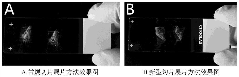
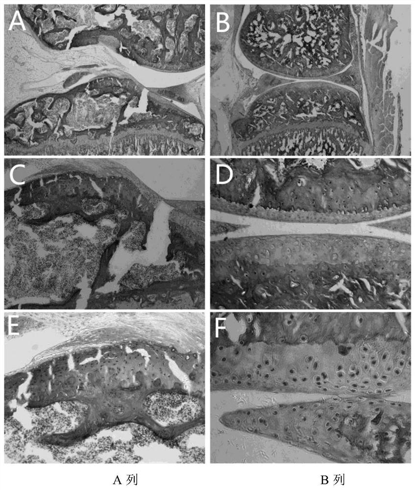
[0040] 4) to attach Figure 4 As an example, a is the paraffin section of the mouse joint tissue processed by the new method of pressing and unfolding, and then HE staining. The results show that the section has no folds, the shape is relatively complete, the layers of each tissue are arranged regularly, the color is bright, and the fine structure inside the cells is clear. , the nucleus c...


PUM

PropertyMeasurementUnit
thicknessaaaaaaaaaa

Abstract

The invention discloses a method and a device for spreading a hard tissue pathological section. The method comprises the following steps: 1) fixing a material, dehydrating, clearing, permeating, embedding and trimming with a grinding wheel; 2) uniformly coating an OCT embedding medium on the surface of the hard section and then slicing; 3) putting the sliced hard tissue section into hot water at 45-52 DEG C and spreading the tissue section; 4) using a glass slide coated with chrome alum gelatin for fishing out the hard tissue section; 5) uniformly coating vaseline on a cover glass and placingon the glass slide adhered to the tissue section; 6) placing the glass slide into a pressing device, uniformly applying a pressure, putting the pressing device with the glass slide into an oven at 60DEG C and baking for 2 hours; and 7) taking the glass slide out of the oven, removing the cover glass, putting into xylene for removing vaseline, and naturally airing for later use. The method and thedevice for spreading the hard tissue pathological section disclosed by the invention have the advantages that the operation is convenient, the repeatability is high, the problems of shrinking, shedding, and the like, of the hard tissue section can be effectively avoided, the spreading effect is obviously promoted and the method and the device are suitable for different tissue-engineered bone researches.

Description

technical field [0001] The invention relates to the technical field of preparation of pathological tissue slices in biology or medicine, in particular to a new method for displaying hard tissue pathological slices. Background technique [0002] Bone tissue is a kind of hard connective tissue composed of cells, matrix and fibers. The inorganic salt deposited in bone tissue exists in the form of hydroxyapatite, and its main components are calcium phosphate and calcium carbonate. Because bone tissue is hard, brittle, dense, difficult to cut and fragile, it is difficult to slice bone tissue directly in clinical, teaching and scientific research. Decalcified and paraffin-embedded sections of conventional bone tissue often lead to decreased enzyme activity and changes in bone tissue morphology and structure during processing, making it impossible to conduct histological observations, and to study the distribution of calcium and mineralization in tissues. In recent years, the rese...

Claims


Application Information

Patent Timeline
Patent Type & Authority Patents(China)
IPC IPC(8): G01N1/28
CPCG01N1/28G01N1/2813G01N1/286
Inventor 雷光华何可邓鑫佳丁翔刘之晨梁旖旎曾超李辉
Owner XIANGYA HOSPITAL CENT SOUTH UNIV
Why Eureka
Try Eureka