Eureka AIR delivers breakthrough ideas for toughest innovation challenges, trusted by R&D personnel around the world.

Rapid detection method of live virus and application of rapid detection method

A detection method and technology for live virus, applied in the field of rapid detection of live virus, to achieve the effect of verifying the effectiveness of virus inactivation process, good quantitative measurement effect, and rapid detection

Inactive Publication Date: 2019-02-15
NAT INST FOR FOOD & DRUG CONTROL
View PDF1 Cites 0 Cited by
  • Summary
  • Abstract
  • Description
  • Claims
  • Application Information

AI Technical Summary

Problems solved by technology

Pseudorabies virus (PRV) is a commonly used indicator virus for virus inactivation verification of animal-derived medical products. At present, there is no rapid detection method for viral nucleic acid PCR that can specifically detect live PRV viruses (not interfered by dead viruses)

Method used


Image

  • Rapid detection method of live virus and application of rapid detection method
  • Rapid detection method of live virus and application of rapid detection method
  • Rapid detection method of live virus and application of rapid detection method

Examples


Embodiment 1

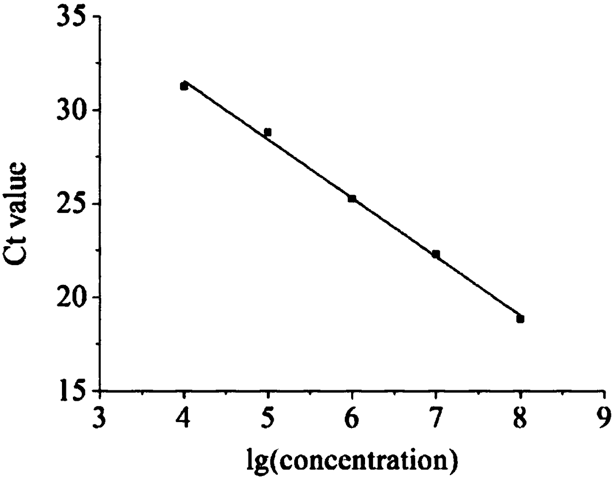
[0061] Establishment and verification of a rapid detection method for live virus (ie, ICC-qPCR method).

[0062] 1. Establishment and verification of qPCR method.

[0063] 1. Preparation of plasmid DNA standards.

[0064] The strain category of PRV (pseudorabies virus) is Barthar strain, and gB is a gene name of this PRV strain. Referring to the gB gene sequence of Barthar strain in GeneBank, after experimental screening and verification, the following primer set was selected as the primers for this experimental research, and the primer sequences are:

[0065] FP: 5'-GTCACCTTGTGGTTGTTG-3' (SEQ ID No. 1)

[0066] RP: 5'-CCACATCTACTACAAGAAC G-3' (SEQ ID No.2)

[0067] The fragment amplified with the above primer set was 180 bp in length. The electrophoresis product of the amplified fragment was recovered and purified, and then connected to the pMDTM18-T cloning vector for cloning. The positive clones were initially identified by nucleic acid electrophoresis and qPCR. The ide...

Embodiment 2

[0113] The application of the ICC-qPCR detection method of PRV in the validity verification of a virus inactivation process (electrophysiological catheter ethylene oxide virus inactivation process).

[0114] 1. Pollutant load.

[0115] Based on the physical characteristics of the electrophysiological catheter, clinical application, and the characteristics of the pollutants, the pollutants in the whole blood test were selected (100 mL of citrated sheep whole blood, 50 mL of calf serum, and 50 mL of normal saline, and 2 mol / L CaCl was added before use). 2 0.01mL), to simulate the contamination after clinical use. A high titer of PRV virus was added to the whole blood test contaminant to prepare the whole blood test contaminant containing PRV virus.

[0116] Using 12 sterile solid electrophysiological catheters, by measuring the length (L) and diameter (2r) of the shaft of the catheter for experimental contamination (measured with a vernier caliper), calculate the approximate s...


PUM


Abstract

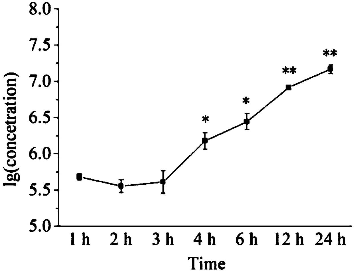
The invention relates to a rapid detection method of live virus and application of the rapid detection method, and belongs to the technical field of biological detection. The rapid detection method comprises the following steps: transfection: inoculating viruses to be detected into single-layer host cells cultured in a cell culture plate in advance, transfecting and culturing; and detection: collecting samples at a sample collecting time when the inoculated viruses to be detected enter the host cells but are not amplified, cleaning for removing the viruses outside the host cells, then extracting viral DNA in the host cells for PCR detection to obtain the content of the live viruses in the viruses to be detected. The rapid detection method can effectively detect residual live viruses aftera virus inactivation process, has a quantification limit of 104 copies.muL-1 (approximately equivalent to 2 lgs) and a detection limit of 103 copies.muL-1 (approximately equivalent to -1 lg) and a minimum detection limit for detecting PRV infection titer of -1 lg, has the sensitivity higher than a traditional CPE method (-0.5 lg), can avoid the influence of subjective factors on judgment of results, and saves time and experiment costs.

Description

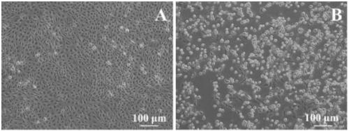
technical field [0001] The invention relates to the technical field of biological detection, in particular to a rapid detection method for live virus and its application. Background technique [0002] The traditional virus titration method based on cytopathic effect (CPE) is a classic method for detecting live viruses, but it often requires serial dilution of samples, and each dilution requires multiple replicate wells to observe the virus infection caused by the microscope. Cytopathic, then calculate the virus per mL log(TCID 50 )number. This method consumes a lot of consumables and observation time for the evaluation of each sample, and the manual observation of cell lesions is easily affected by subjective factors. In addition, this method usually requires long-term cultivation to quantify infectious viruses, especially for viruses that develop slowly. Due to the long culture time, the cells themselves may change when no cytopathic changes occur, and it is impossible to...

Claims


Application Information

Patent Timeline
IPC IPC(8): C12Q1/70C12Q1/6851C12R1/93
CPCC12Q1/6851C12Q1/705C12Q2531/113C12Q2545/114
Inventor 徐丽明魏慧慧段晓杰张羽陈亮邵安良魏利娜张乐李天任王玉梅
Owner NAT INST FOR FOOD & DRUG CONTROL
Why Eureka
Eureka Blog
Learn More