Supercharge Your Innovation With Domain-Expert AI Agents!

Method for carrying out plant tissue fluorescent microscopic analysis by organic polymer/silicon dioxide composite nano film

A plant tissue and analysis method technology, which is applied in the field of fluorescence microscopic imaging analysis, can solve the problems of limited application, and achieve the effect of simple method and low cost

Active Publication Date: 2019-04-09
QILU UNIV OF TECH
View PDF9 Cites 0 Cited by
  • Summary
  • Abstract
  • Description
  • Claims
  • Application Information

AI Technical Summary

Problems solved by technology

However, the relative quantum yield of most carbon-based fluorescent materials does not exceed 50% (ACS applied materials & interfaces, 7(2015) 23231-23238; ACS nano, 9(2015) 11455-11461.), thus limiting their applications

Method used


Image

  • Method for carrying out plant tissue fluorescent microscopic analysis by organic polymer/silicon dioxide composite nano film
  • Method for carrying out plant tissue fluorescent microscopic analysis by organic polymer/silicon dioxide composite nano film
  • Method for carrying out plant tissue fluorescent microscopic analysis by organic polymer/silicon dioxide composite nano film

Examples


Embodiment 1

[0046] A preparation method of organic polymer / silicon dioxide composite nano film, the steps are as follows:

[0047] (1) Mix citric acid and L-cysteine ​​at a ratio of 2:1, grind evenly, then transfer to a beaker, heat the beaker in an oven at 160°C for 1 hour; take the beaker out and place it at room temperature Cool in the environment to obtain brown yellow powder A.

[0048] (2) Weigh 65mg of powder A, dissolve it in 58.5mL of distilled water with the assistance of ultrasound (100W, 40kHz, 5min), and obtain aqueous solution B of fluorescent organic polymer nanofilm; In absolute ethanol, solution C was obtained.

[0049](3) Under stirring, solution C was added to solution B, stirred and reacted at room temperature for 12 hours, and mixed solution D was obtained.

[0050] (4) Freeze-dry the mixed solution D to obtain an organic polymer / silicon dioxide composite nano-membrane powder, which is designated as sample S-1.

Embodiment 2

[0052] The 5.4 μmol of ethyl orthosilicate in step (2) of Example 1 was replaced with 21.6 μmol of ethyl orthosilicate, and the other conditions remained unchanged.

[0053] The obtained organic polymer / silicon dioxide composite nano-membrane powder sample is designated as S-2.

Embodiment 3

[0055] The 5.4 μmol orthoethyl silicate in the step (2) of Example 1 was replaced with 43.2 μmol orthoethyl silicate, and the other conditions remained unchanged.

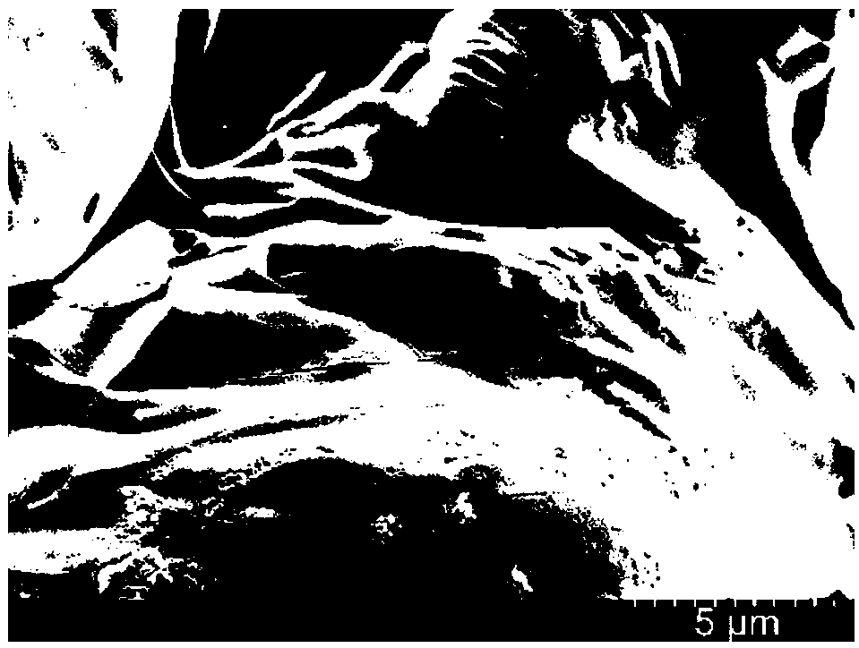
[0056] Gained organic polymer / silicon dioxide composite nano-film powder sample is denoted as S-3, and sample S-3 is taken for SEM observation (attached figure 1 ).


PUM


Abstract

The invention relates to a plant tissue fluorescent microscopic imaging analysis method, in particular to a fluorescent microscopic imaging analysis method using a fluorescent organic polymer / silicondioxide composite nano film. According to the plant tissue fluorescent microscopic imaging analysis method, the organic polymer / silicon dioxide composite nano film is used; and 0.1-1 mg / mL nano film water solution is used for culturing biological tissues. The obtained organic polymer / silicon dioxide composite nano film has biocompatibility, can be used as culture solution for biological tissues, and can be used as a fluorescent indicator for biological tissue fluorescent microscopic imaging.

Description

technical field [0001] The invention relates to a fluorescent microscopic imaging analysis method of plant tissue, in particular to a fluorescent microscopic imaging analysis method using a fluorescent organic polymer / silicon dioxide composite nano film. Background technique [0002] Observation experiments on plant tissues often use the slice observation method. Although slice observation can conveniently observe the morphology of plant tissues, it will inevitably damage plant tissues and is not suitable for in vivo analysis. [0003] Biocompatible carbon-based fluorescent materials have important applications in the fields of sensing, cell imaging, and cancer diagnosis and treatment (Sensors and Actuators B: Chemical, 242 (2017) 1210-1215; ACS applied materials & interfaces, 7 (2015) 23231 -23238; ACS nano, 9(2015) 11455-11461.). If it is used for the fluorescence analysis of living organisms, the concentration of the fluorescent material should be as low as possible; th...

Claims


Application Information

Patent Timeline
Patent Type & Authority Applications(China)
IPC IPC(8): G01N21/64G01N1/28
CPCG01N1/28G01N21/6402
Inventor 班青盖利刚冯宝羲
Owner QILU UNIV OF TECH
Features
  • R&D
  • Intellectual Property
  • Life Sciences
  • Materials
  • Tech Scout
  • Unparalleled Data Quality
  • Higher Quality Content
  • 60% Fewer Hallucinations
Learn More