
Human glioma primary cells and in-vitro isolation and culture method and application thereof

A primary cell, isolation and culture technology, applied in the field of cell biology, can solve the problem that the real physiological response of clinical patients cannot be accurately reflected, the low-grade glioma-related research cannot provide an effective research system, and the loss of primary glioma cells can be solved. Heterogeneity and other issues to achieve the effect of stable traits

Active Publication Date: 2019-04-30
武汉赛尔朗灵科技有限公司
View PDF2 Cites 3 Cited by
  • Summary
  • Abstract
  • Description
  • Claims
  • Application Information

AI Technical Summary

Problems solved by technology

This application involves a high-grade glioma cell line. Previous studies have believed that the morphology of cell lines with multiple passages in vitro tends to be uniform, and gradually loses the heterogeneity of primary glioma cells. Due to changes in their genetic background, it is impossible to accurately Reflect the real physiological response of clinical patients
Moreover, this application only involves high-grade cell lines of glioma, and cannot provide an effective research system for related research on low-grade glioma

Method used


Image

  • Human glioma primary cells and in-vitro isolation and culture method and application thereof
  • Human glioma primary cells and in-vitro isolation and culture method and application thereof
  • Human glioma primary cells and in-vitro isolation and culture method and application thereof

Examples


Embodiment 1

[0038] Example 1: Primary isolation and culture of human high-grade glioma primary cells

[0039] (1) Through the hospital ethics committee, with the consent of the patient or the patient's guardian and after signing the informed consent, fresh clinical glioma resection specimens were obtained from Wuhan Union Medical College Hospital. The specimens were WHO grade glioblastoma.

[0040] (2) Immediately put the resected specimen into the pre-cooled sterile tissue preservation solution (containing 1000U / ml penicillin, 1000ug / ml streptomycin sulfate, 2.5ug / ml amphotericin and 50ug / ml gentamicin) Into the collection tube, and immediately put into a 4 ℃ sample transport box, transported to the laboratory within 4 hours for cell separation.

[0041] (3) Primary isolation culture: Obtain the tissue in a biological safety cabinet, rinse it once with absolute ethanol, and rinse it twice with 1xPBS (pH7.2-7.4), remove blood vessels, fat and For the necrotic tissue part, cut the tissue ...

Embodiment 2

[0043] Example 2: Primary isolation and culture of primary human low-grade glioma cells

[0044] (1) Through the hospital ethics committee, with the consent of the patient or the patient's guardian and signed the informed consent, a fresh clinical glioma resection specimen was obtained from Wuhan Union Medical College Hospital. The specimen was WHO grade II diffuse astrocytoma.

[0045] (2) Immediately put the resected specimen into the pre-cooled sterile tissue preservation solution (containing 1000U / ml penicillin, 1000ug / ml streptomycin sulfate, 2.5ug / ml amphotericin and 50ug / ml gentamicin) Into the collection tube, and immediately put into a 4 ℃ sample transport box, transported to the laboratory within 4 hours for cell separation.

[0046] (3) Primary isolation culture: Obtain the tissue in a biological safety cabinet, rinse it once with absolute ethanol, and rinse it twice with 1xPBS (pH7.2-7.4), remove blood vessels, fat and For the necrotic tissue part, cut the tissue ...

Embodiment 3

[0048] Example 3: Subculture of primary human glioma cells

[0049] (1) When the abundance of cells cultured in T25 culture flask reaches 80%, rinse the cells twice with 1xPBS (pH7.2-7.4), and add 1ml of 0.05% trypsin-EDTA to digest the monolayer cells for 2-3min.

[0050] (2) Add 2ml DF31 complete medium to stop the digestion.

[0051] (3) Centrifuge at 1000rpm for 4 minutes, remove the supernatant, collect the cell suspension, resuspend with 1ml DF31 complete medium, supplement the medium, and put it into a T25 culture bottle for cultivation according to the ratio of 1 to 2.

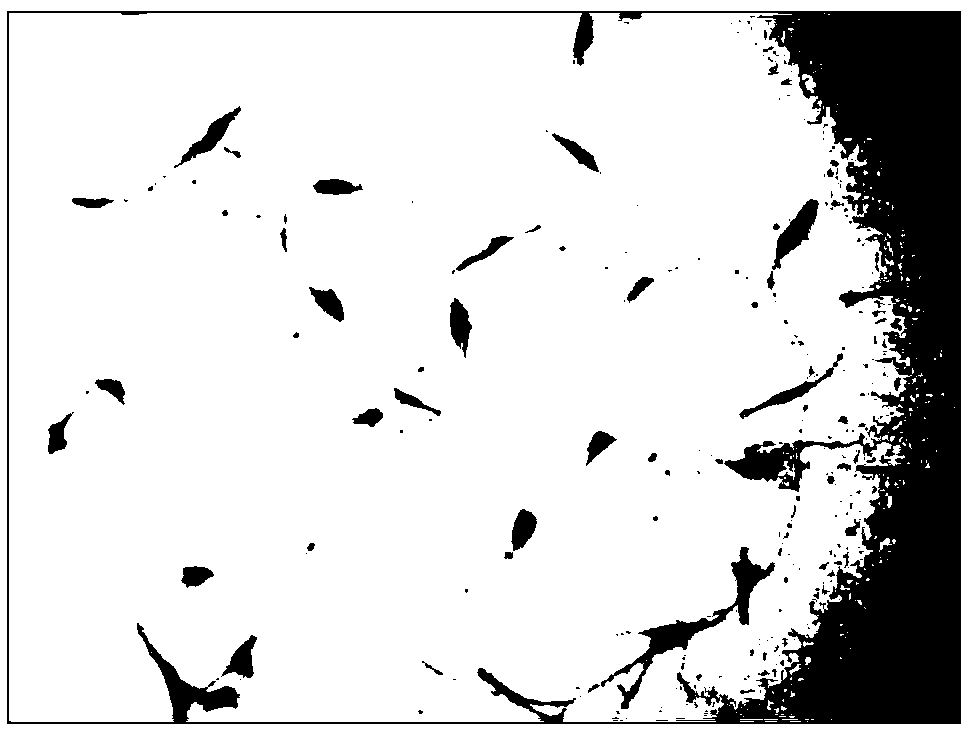
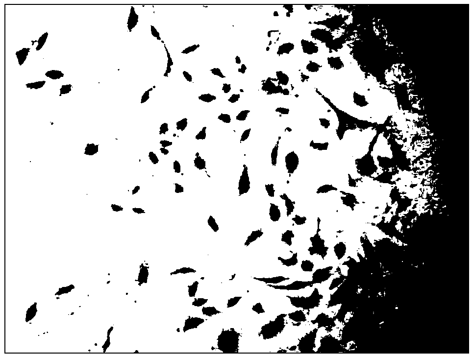
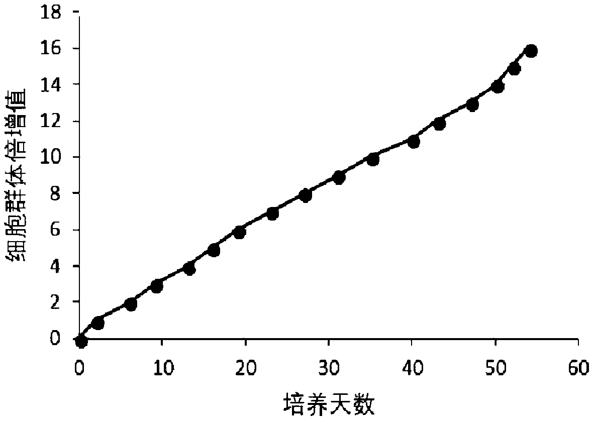
[0052] The human high-grade glioma primary cells and human low-grade glioma primary cells subcultured according to the above method, the cell growth curve of the culture establishment line is as follows: image 3 with Figure 4 , continuous passage for 50 days, the primary human glioma cells of the present invention can still maintain a proliferative state and grow normally.


PUM


Abstract

The invention discloses human glioma primary cells and an in-vitro isolation and culture method and an application thereof. The cells are named as a low-grade human glioma primary cell XHLG-07 and a high-grade human glioma primary cell XHHG-02 and have the preservation numbers of CCTCC NO:C2018256 and CCTCC NO:C2018215 respectively. The isolation and culture method comprises the steps: acquiring aglioma tissue excised after operation in clinic in vitro, removing blood vessels in the tissue and an adipose tissue by anatomical tweezers, digesting by collagenase / pancreatin, filtering, carrying out low-speed centrifugation to obtain a cell precipitate, carrying out lysis of erythrocytes with an erythrocyte lysis solution, resuspending cells in a DF31 complete culture medium and carrying out inoculation culture. The isolation method can be used for acquiring WHO glioma primary cells with different grades. The obtained cells can be used for analyzing pathogenesis, drug sensitivity and metastasis of human brain gliomas with different grades in vitro and in vivo, and provide research materials more similar to the biological characteristics of clinical tumors for the study of human brain gliomas with different grades.

Description

technical field [0001] The invention belongs to the field of cell biology, and more specifically relates to a primary human glioma cell and its in vitro separation and culture method and application. Background technique [0002] Glioma is the most common intracranial malignant tumor of the nervous system, which originates from glial cells. According to the WHO classification of central nervous system tumors, gliomas are divided into WHO I-IV grades, in which I and II are low-grade gliomas, and III and IV are high-grade gliomas. In the past 30 years, the incidence of primary malignant brain tumors has been increasing year by year, especially in the elderly. The incidence of glioma accounts for about 30% of all central nervous system tumors, among which glioblastoma has the highest incidence rate of about 3.2 / 100,000, followed by diffuse astrocytoma with an incidence rate of 0.53 / 100,000 . Despite the application of various treatment methods, including surgical resection, ...

Claims


Application Information

Patent Timeline
Patent Type & Authority Applications(China)
IPC IPC(8): C12N5/09C12Q1/02
CPCC12N5/0693C12N2500/12C12N2500/24C12N2500/30C12N2500/32C12N2501/11C12N2501/115C12N2501/392C12N2501/91C12N2503/02G01N33/5011G01N2500/10
Inventor 刘红亚熊南翔李俊俊王前进王轩徐豪王艺淏屈彦魁
Owner 武汉赛尔朗灵科技有限公司
Learn More