Anti-H7N9 whole human monoclonal antibody 5G22, and preparation method and application thereof

A monoclonal antibody, fully human technology, applied in the field of immunology, can solve the problems of rimantadine drug resistance and no effective treatment methods, and achieve the goal of reducing cumbersome operations and costs, high affinity and specificity, and low production costs Effect

Active Publication Date: 2019-07-02
SHENZHEN INST OF ADVANCED TECH CHINESE ACAD OF SCI
4 Cites 0 Cited by

AI-Extracted Technical Summary

Problems solved by technology

H7N9 virus is a kind of influenza virus, which is resistant to the traditional antiv...

Abstract

The present invention relates to an anti-H7N9 whole human monoclonal antibody 5G22, and a preparation method and an application thereof, the anti-H7N9 whole human monoclonal antibody 5G22 or a biologically active fragment derived from the monoclonal antibody and capable of specifically binding H7N9. The antibody has a heavy chain variable region and a light chain variable region. The heavy chain variable region and light chain variable region respectilvey have 3 complementarity determining regions (CDRs). The antibody 5G22 is capable of specifically binding, detecting and neutralizing H7N9 viruses, and more suitable and more promising as a macromolecular drug for treating influenza viruses.

Application Domain

Immunoglobulins against virusesAntivirals +6

Technology Topic

Macromolecular drugHeavy chain +5

Image

  • Anti-H7N9 whole human monoclonal antibody 5G22, and preparation method and application thereof
  • Anti-H7N9 whole human monoclonal antibody 5G22, and preparation method and application thereof
  • Anti-H7N9 whole human monoclonal antibody 5G22, and preparation method and application thereof

Examples

  • Experimental program(3)

Example Embodiment

[0042] Example 1
[0043] (1) Construction of NTH-3T3 cell line stably expressing CD40L
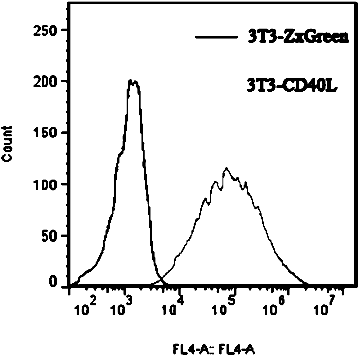
[0044] Use lentivirus to establish 3T3-CD40L feeder cells. The lentiviral expression vector pLVX-CD40L was constructed and transfected into 293T cells. The viral supernatant was collected on the fourth day of transfection. Activated NIH-3T3 cells, cultured for 3 generations and then infected with lentivirus, continued to culture and passage 3 times. Use flow cytometry to sort cells with FITC fluorescence intensity near MFI, and re-add them to the culture flask at 37°C, 5% CO 2 Culture and test in an incubator, the test results are as follows figure 1 As shown, 3T3 cells expressing CD40L and 3T3 cells transfected with empty vector pLVX (with ZxGreen) were stained with anti-CD40L with APC, and then analyzed by flow cytometry. It was found that all 3T3-CD40L feeder cells expressed CD40L. When the cells grow to 80%~90%, digest and collect the cells at a concentration of 1×10 per ml 7 cell. Place in a radiometer for 5000rads radiation, and resuspend the cells in cryopreservation solution at a concentration of 3.5×10 per ml 7 Cells, 1ml aliquoted into cryovials, frozen in liquid nitrogen (can be stored for 2 years).
[0045] (2) Sorting and activation of memory B cells
[0046] Separate and freeze the PBMC of recovered patients who have been infected with H7N9 virus with lymphatic separator, 10~50×10 per tube 6 Cells are frozen in liquid nitrogen tanks. Prepare PBMC flow dyeing solution, its components are shown in Table 1 below:
[0047] Table 1: PBMC flow dyeing solution
[0048] antibody Volume (μL) CD19-PE-Cy7 0.5 IgM-PE 1.0 IgA-APC 2.5 IgD-FITC 2.5 PBS-1%(wt/vol)BSA 43.5
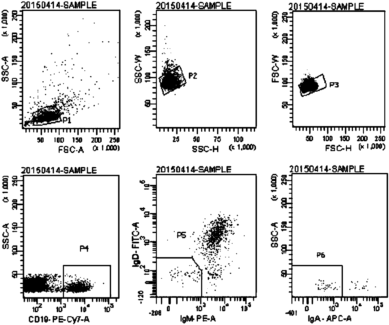
[0049] Thaw PBMC, add the above PBMC flow cytometry staining solution and sort on the flow cytometer, the results are as follows figure 2 As shown, CD19 is sorted out + IgM - IgA - IgD - For memory B cells, the cell purity needs to be above 90%. If it is less than 90%, repeat the sorting process.
[0050] Prepare a mixed medium for activating B cells, the components of which are shown in Table 2 below:
[0051] Table 2
[0052] Component volume Complete IMDM medium 336mL IL-2(10,000U mL -1 )
[0053] Add memory B cells to the mixed medium, mix well and limit dilution in a 384-well plate, with 1 cell per well, with a volume of 50ul, at 37°C, 5% CO 2 Let stand in the incubator for cultivation. After 13 days, the supernatant was taken for ELISA.
[0054] (3) Obtain the human monoclonal antibody 5G22 against H7N9 virus
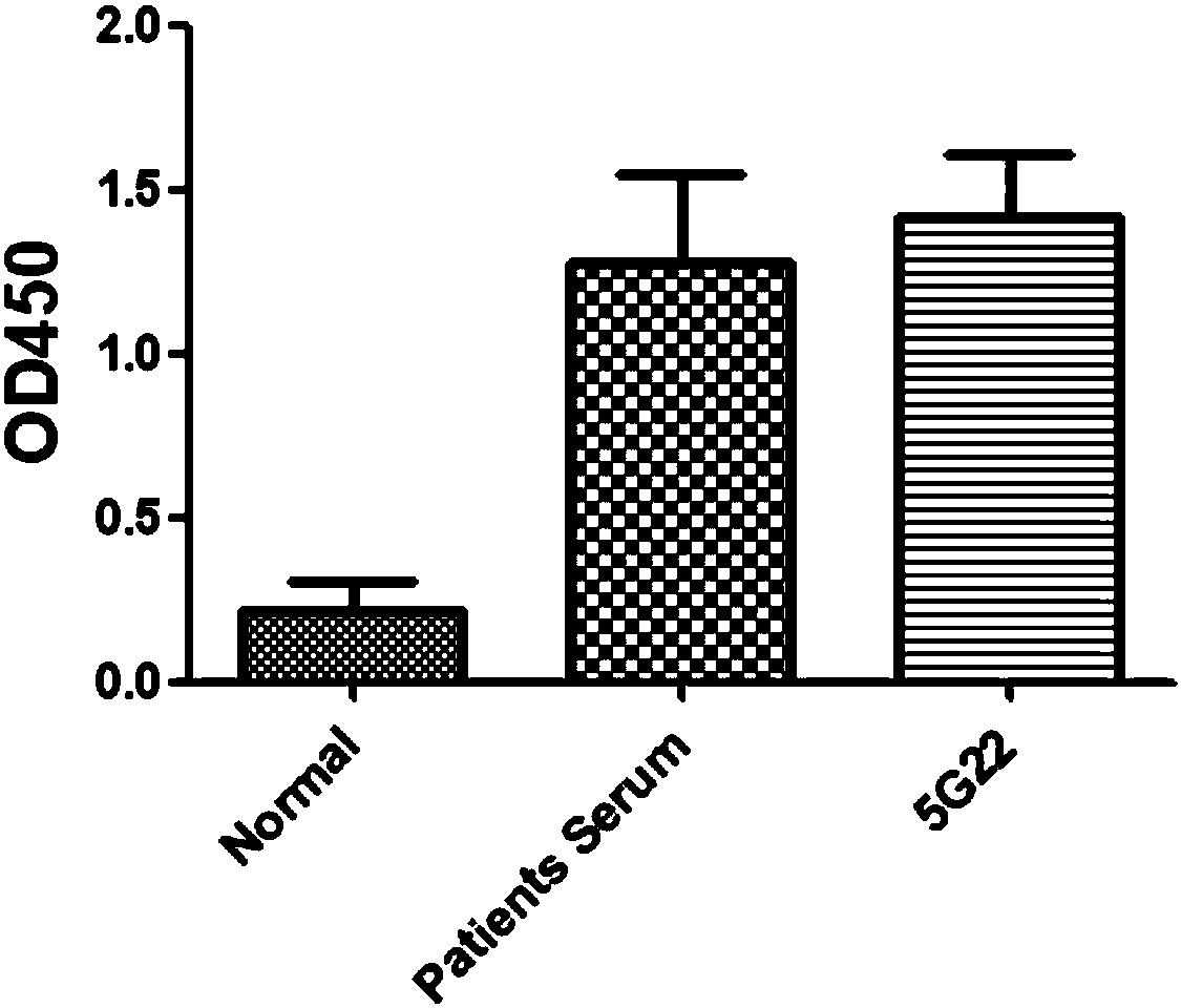
[0055] Influenza virus hemagglutinin HA is a columnar antigen on the surface of the virus envelope. It can bind to a variety of red blood cell receptors such as humans, chickens, and guinea pigs to cause red blood cell agglutination. It is immunogenic. Anti-hemagglutinin antibodies can neutralize influenza viruses. In the present invention, B cells capable of secreting the antibody 5G22 that binds to the H7N9 virus were discovered by ELISA, and the human monoclonal antibody 5G22 secreted by them can target the hemagglutinin HA ( image 3 ).
[0056] Specific operation of ELISA experiment:
[0057] (1) Coat 100ng/100μl of H7N9 virus HA protein in a 96-well microtiter plate, 100μl per well;
[0058] (2) Place in a refrigerator at 4 degrees overnight;
[0059] (3) Wash three times with PBST solution, add 200μl of 5% skimmed milk powder solution to each well, incubate at 37°C for 1 hour;
[0060] (4) Wash three times with PBST solution, add 100μl of normal human serum (negative control) without virus infection, or add virus-infected patient serum or anti-H7N9 fully human monoclonal antibody, three times for each;
[0061] (5) After incubating at 37°C for 1 hour, wash with PBST solution three times;
[0062] (6) Dilute the anti-human IgG antibody with HRP at 1:5000, add it to the enzyme-labeled plate, 100μl per well;
[0063] (7) After incubating at 37°C for 1 hour, wash with PBST solution three times;
[0064] (8) Add 100μl TMB substrate solution to each well, 37°C for 5 minutes;
[0065] (9) Add 100μl of stop solution 2M sulfuric acid to each well, and immediately detect the absorbance at 450nm wavelength in the microplate reader. Such as image 3 As shown, the ELISA experiment shows that the human monoclonal antibody 5G22 obtained in the present invention can target the hemagglutinin HA of H7N9 virus.

Example Embodiment

[0066] Example 2 Cloning, recombination and expression of humanized monoclonal antibody 5G22 gene
[0067] The B cells that can secrete the antibody 5G22 that binds to the H7N9 virus obtained in Example 1 were lysed, and the lysate was taken for reverse transcription of RNA to obtain the PCR template cDNA of the human antibody gene. The heavy chain and light chain genes of the antibody were cloned using cDNA as a template, and recombined in eukaryotic cells 293F or HEK293 for expression and purification.
[0068] specifically:
[0069] (1) Transfer the B cell liquid to a 96-well plate (Eppendorf, 030133366).
[0070] (2) Reverse transcription system: 150ng random primer (invitrogen, 48190-011), 0.5ul 10mM dNTP (Invitrogen, 18427-088), 1μl 0.1M DTT (Invitrogen, 18080-044), 0.5%v/v Igepal CA -630 (Sigma, I3021-50ML), 4U RNAsin (Promega), 6U Prime RNAse Inhibitor
[0071] (Eppendorf)and 50U III reverse transcriptase (Invitrogen, 18080-044), supplement with DEPC water to 14μl/well.
[0072] (3) Reverse transcription reaction program: 42°C, 10min; 25°C, 10min; 50°C, 60min; 94°C, 5min.
[0073] (4) cDNA is stored at -20°C.
[0074] (5) Use KOD-Plus-Neo (TOYOBO, KOD401) kit PCR to amplify the heavy and light chains of antibody genes, 40μL system: 3.5μL cDNA, 20nM mixed primers, 4μL buffer, 4μL 2mM dNTPs, 2.4μLMgSO 4 , 1μL KOD.
[0075] (6) Reaction procedure: 94℃, 2min; 45 cycles: [98℃, 10s; 58℃ (IgH/Igκ) or 60℃ (Igλ), 30s; 68℃, 28s (1 st PCR) or 23s(2 nd PCR)].
[0076] (7) Perform agarose gel on the amplified product, and the result is as follows Figure 4 As shown, the results show that the variable region of the light chain of the antibody is κ with a size of 321 bp, and the size of the variable region of the heavy chain is 366 bp.
[0077] (8) The sequencing results of antibody gene PCR products are as follows:
[0078] 5G22 heavy chain variable region gene (SEQ ID NO:1):
[0079] GAGGTGCAGCTGGTGGAGTCTGGGGGAGGCTTGGCCCAGCCTGGGGGGTCCCTGAGACTCTCCTGTGCAGCCTCTGGATTCAGCTTTAGTACCTATTGGATGACCTGGGTCCGCCAGGCTCCAGGGAAGGGGTTGGAGTGGGTGGCCAACATAAAGCAAGACGGAGGTGAGAAATACTATGTGGACTCTGTGAAGGGCCGATTCATCGTCTCCAGAGACAACGCCCAGAACTCACTGGATCTGCAAATGAACAGCCTGAGGGCCGAGGACACGGCTGTGTATTACTGTGCCAGAGATTCAGGCACGTTTCTTTGGAGTGGTTATCCGGACTACTGGGGCCAGGGAACCCTGGTCACCGTCTCCTCA
[0080] The light chain variable region gene of 5G22 (SEQ ID NO: 3):
[0081] GacatccagATGACCCAGTCTCCATCCTCCCTGTCTGCATCTGTACGAGACAGAGTCACCATCACTTGCCGGGCCAGTCAAAATATTAATAGCTGGTTGGCCTGGTATCAGCAGAAACCAGGGAAAGCCCCTAAACTCCTGATCTATAAGGCGTCTAGTTTAGAAAGTGGGGTCCCATCAAGGTTCAGCGGCAGTGGATCTGGGACAGAATTCACTCTCACCATCAGCAGCCTGCAGCCTGATGATTTTGCAACTTATTACTGCCAACAGTATAAAAATTATTCTCGGACGTTCGGCCAAGGGACCAAGGTGGAAATCAAA
[0082] (9) The H gene and pcDNA3.1 were respectively subjected to BamH1/EcoR1 double enzyme digestion and then connected to form a pcDNA3.1-H vector.
[0083] (10) The L gene and pcDNA3.1 were respectively subjected to Not1/Xho1 double enzyme digestion and then connected to form a pcDNA3.1-L vector.
[0084] (11) Culture 293F cells.
[0085] (12) 20ug pcDNA3.1-L vector and 10ug pcDNA3.1-H vector were co-transfected into 293F cells and cultured for 96 hours.
[0086] (13) Take the supernatant and perform ELISA (ABC is the supernatant, DEF is the positive control, and GH is the negative control) and western blot; the results of the ELISA experiment are shown in Table 3 below:
[0087] table 3
[0088] data 450 data 450 A 1.4025 E 1.2587 B 1.4215 F 1.1247 C 1.3956 G 0.1655 D 1.3563 H 0.1741
[0089] The above results show that the supernatant contains the antibody 5G22 capable of binding to the H7N9 virus.
[0090] The specific process of Western blot experiment is:
[0091] Run protein denaturation electrophoresis with the supernatant, block with 5% skimmed milk powder solution for 1 hour after transfer, and then incubate with goat anti-human IgG antibody with HRP for 1 hour, and finally add display substrate for exposure. The experimental results are as Figure 4 As shown, Figure 4 The heavy and light chains of the fully human antibody are shown, indicating that the supernatant contains the fully human monoclonal antibody 5G22 against H7N9 virus.
[0092] For the monoclonal antibody 5G22 of this embodiment, SEQ ID NO: 2 and SEQ ID NO: 4 are the VH and VL amino acid sequences of the heavy chain variable region and the light chain variable region of 5G22 obtained in this embodiment, respectively. See Figure 5 , The heavy chain variable region and the light chain variable region each have 3 complementarity determining regions (CDR), respectively numbered CDR1, CDR2, CDR3, where: heavy chain: CDR1: GFSFSTYW (SEQ ID NO: 5); CDR2: IKQDGGEK (SEQ ID NO: 6); CDR3: ARDSGTFLWSGYPDY (SEQ ID NO: 7). Light chain: CDR1: QNINSW (SEQ ID NO: 8); CDR2: KAS; CDR3: QQYKNYSRT (SEQ ID NO: 9).

Example Embodiment

[0093] Example 3 In vitro neutralization experiment of humanized monoclonal antibody 5G22
[0094] Using a virus-infected cell model (canine kidney cell MDCK), the inhibitory effect and effect of the antibody on the H7N9 influenza virus with PR-8 as the skeleton were evaluated by the micro-neutralization-ELISA experiment, and the anti-influenza virus activity of the antibody was detected. The specific operations are as follows:
[0095] 1 cell plating
[0096] Trypsin digests the cells in logarithmic growth phase, collect them by centrifugation after termination, blow evenly to prepare single cell suspension;
[0097] Adjust the cell concentration to 5×10⁴ cells/ml with cell culture solution, inoculate it in a 96-well cell culture plate, and place the cells at 37°C, 5% CO 2 Cultivate overnight in an incubator.
[0098] 2 Antibody and virus pretreatment
[0099] 5G22 antibody has 10 concentration gradients, 10-10 10 Double dilution, each group has 3 parallel holes for each concentration.
[0100] H7N9 virus A/Anhui/1/2013 was taken from the Institute of Microbiology, Chinese Academy of Sciences.
[0101] 3 Virus infection
[0102] Discard the cell culture supernatant and wash 3 times with PBS. Put the premixed antibody-virus mixture (in 10 2 μg/ml of 5G22 monoclonal antibody is 10-10 10 Double dilution, the 5G22 antibody of each concentration was mixed with an equal volume of 100TCID 50 The virus was mixed to obtain the mixture) was added to a 96-well cell culture plate, incubated at 37°C for 1 hour, aspirated and discarded, and washed twice with PBS.
[0103] 4 Configure maintenance fluid
[0104] Add TPCK-Trypsin with a final concentration of 2μg/ml to DMEM without serum with double antibody. Discard the PBS in the 96-well plate, add 100μl maintenance solution to each well, and place at 37°C, 5% CO 2 Incubate for 20h in the incubator.
[0105] 5 Neutralization experiment-ELISA method
[0106] (1) Discard the cell fluid in the micro culture plate;
[0107] (2) Wash the cells once with 100μl PBS;
[0108] (3) Discard PBS (don't let the cells dry), and add 50μl/well of fixative (volume ratio acetone: absolute ethanol = 2:3);
[0109] (4) Cover the micro culture plate and fix the cells at room temperature for 10 minutes;
[0110] (5) Discard the fixative solution, wash the cells with 100 μl PBS solution, and repeat the washing 3 times (shake gently to avoid strong washing) to remove residual acetone.
[0111] (6) Block the cells with 5% skimmed milk powder at room temperature for 1 hour, and wash the cells with 100 μl PBS solution once;
[0112] (7) Dilute 1 antibody (anti-influenza virus nucleoprotein-NP monoclonal antibody, commercially available) with PBS 1:2000, add 50 μl of the diluted solution to each well, and react for 1 hour at room temperature.
[0113] (8) Wash the plate 5 times with 100μl PBST to remove 1 antibody;
[0114] (9) Dilute 2 antibody (anti-mouse IgG antibody with HRP) in PBS 1:2000, add 50μl to each well, and act at room temperature for 1 hour;
[0115] (10) Wash the plate 6 times with 100μl PBST to remove 2 antibodies;
[0116] (11) Add 50μl of TMB color developing solution to each well;
[0117] (12) After 10 minutes of color development at room temperature in the dark, add 50μl of 2M hydrochloric acid to each well to stop the reaction;
[0118] (13) Read the OD value of each well on the ELISA tester (450 nm).
[0119] Statistical Analysis
[0120] Use GraphPad Prism 6.0.1 to analyze the data and draw a dose-response curve, and calculate the IC50. Inhibition rate calculation formula: Inhibition rate=[(OD virus well-OD negative cell control well)-(OD drug well-OD negative cell control well)]/(OD virus well-OD negative cell control well)×100%.
[0121] Experimental results
[0122] The result is as Image 6 As shown, the 5G22 of the present invention has good broad-spectrum neutralization.

PUM

no PUM

Description & Claims & Application Information

We can also present the details of the Description, Claims and Application information to help users get a comprehensive understanding of the technical details of the patent, such as background art, summary of invention, brief description of drawings, description of embodiments, and other original content. On the other hand, users can also determine the specific scope of protection of the technology through the list of claims; as well as understand the changes in the life cycle of the technology with the presentation of the patent timeline. Login to view more.

Similar technology patents

Graft and rapid propagation method for commercial production of Paeonia rockii

InactiveCN101473741AShorten the out-of-the-garden cyclereduce manufacturing cost
Owner:兰州牡丹园艺开发公司

Classification and recommendation of technical efficacy words

  • Easy to operate
  • reduce manufacturing cost

Lancet device

InactiveUS7303573B2inexpensively mass-producedeasy to operate
Owner:ABBOTT DIABETES CARE INC

Rapid and non-invasive optical detection of internal bleeding

InactiveUS20050065436A1easy to operaterapid and accurate result
Owner:HO WINSTON ZONH +2
Why Eureka
Try Eureka