
Method of detecting extracellular vesicle surface PD-L1 protein by aptamer of programmed death receptor-ligand 1 (PD-L1)

A PD-L1 and programmed death technology is applied in the field of the detection of PD-L1 protein on the surface of extracellular vesicles by nucleic acid aptamer of programmed death receptor-ligand 1, which can solve the problem of cumbersome steps, complicated and time-consuming detection methods. , large sample consumption, etc., to achieve the effect of economical price, fast and efficient detection, and high sensitivity

Active Publication Date: 2019-08-09
XIAMEN UNIV
View PDF1 Cites 6 Cited by
  • Summary
  • Abstract
  • Description
  • Claims
  • Application Information

AI Technical Summary

Problems solved by technology

[0010] The main purpose of the present invention is to overcome the uncertainty and incompleteness of detecting the expression level of tumor cell PD-L1 in the prior art, as well as the problems of cumbersome detection method steps, complex and time-consuming operation, large sample consumption, and high price. Provides a method for detecting PD-L1 protein on the surface of extracellular vesicles with a nucleic acid aptamer for programmed death receptor-ligand 1, using a nucleic acid aptamer that recognizes PD-L1 with high specificity and high affinity to detect extracellular vesicles The PD-L1 expressed on the surface can be detected by micro-thermophoresis equipment. This method is simple, fast, efficient, affordable, sensitive and sensitive, and consumes a small amount of detection samples.

Method used


Image

  • Method of detecting extracellular vesicle surface PD-L1 protein by aptamer of programmed death receptor-ligand 1 (PD-L1)
  • Method of detecting extracellular vesicle surface PD-L1 protein by aptamer of programmed death receptor-ligand 1 (PD-L1)
  • Method of detecting extracellular vesicle surface PD-L1 protein by aptamer of programmed death receptor-ligand 1 (PD-L1)

Examples


Embodiment 1

[0035] Example 1 Extraction of exosomes secreted by melanoma cell line A375 by differential centrifugation

[0036] For the exosomes extracted from the culture supernatant of the melanoma cell line A375 cells, avoid the interference from the exosomes in the fetal bovine serum, by centrifuging the fetal bovine serum overnight at 100000×g to remove the exosomes, and obtain bovine-free exosomes. Fetal Bovine Serum (FBS). Then, the cells were cultured at 37°C in a medium supplemented with 10% exosomal depleted FBS and 1% (v / v) penicillin-streptomycin. After 60 hours of incubation, collect the cell culture supernatant and centrifuge at 2000×g for 20 minutes at 4°C (Eppendorf, 5424R) to remove cells and cell debris. Collect the supernatant and centrifuge at 16500×g for 45 minutes at 4°C To remove a large number of microvesicles (Eppendorf, 5424R). The microvesicle pellet was resuspended in PBS and stored in a refrigerator at 4°C. The supernatant was centrifuged at 100000g for 2 hour...

Embodiment 2

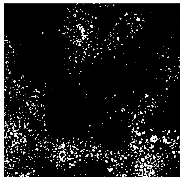
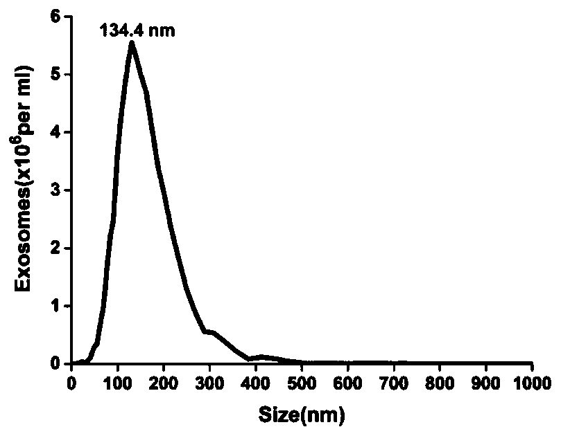
[0037] Example 2 Morphological characterization of exosomes secreted by melanoma cell line A375

[0038] In order to identify exosomes, morphological analysis of exosomes is required. Then, in order to characterize the purified small vesicles as exosomes, they can be characterized by transmission electron microscopy and nanoparticle tracking analyzer. First, the morphology and size of the exosomes are characterized by transmission electron microscopy. The specific operation: the exosomes are dropped on the carbon-coated copper grid and left for 1 minute (the copper grid should be lightly clamped with tweezers to prevent the copper grid Clipped), after staining with 2% uranyl acetate at room temperature for 1 minute, blot the dye solution with filter paper along the edges, put the copper mesh under a lamp and bake for 10 minutes, use a transmission electron microscope (Tecnai, G2spirit FEI, USA) Observe and take pictures. The concentration and particle size distribution of exoso...

Embodiment 3

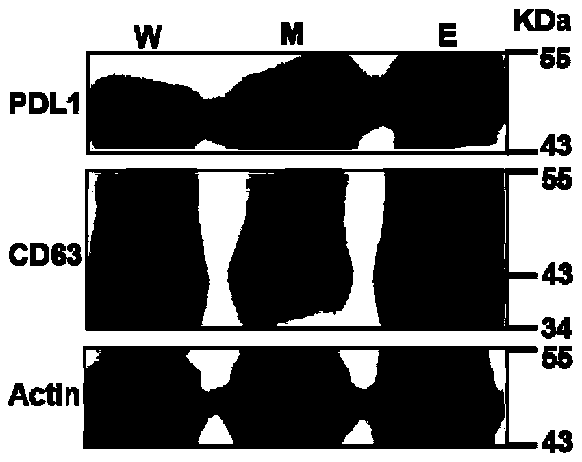
[0040] Example 3 Verification of the expression of PD-L1 in exosomes secreted by the melanoma cell line A375

[0041] After successfully extracting exosomes, it is important to characterize the proteins expressed by exosomes, such as CD63 and PD-L1, which can be identified by Western blotting. Using 12% SDS-PAGE electrophoresis analysis, load cell lysate (W) microvesicles (M) and exosomes (E) respectively, load the same total amount of protein, and run three gels in parallel; then transfer to nitric acid On the cellulose membrane, the blot was blocked with blocking buffer (Beyotime Biotechnology) for 1 hour at room temperature. After washing, it was incubated with PD-L1, CD63 and Actin primary antibodies at 4°C overnight. After washing, it was conjugated with HRP The secondary antibody (Cell Signaling Technology) was incubated together at room temperature for 2 hours; after washing, the blot on the membrane was developed with ECL detection reagent (Pierce), and the chemiluminesce...


PUM

PropertyMeasurementUnit
The average particle sizeaaaaaaaaaa

Abstract

The invention discloses a method of detecting an extracellular vesicle surface PD-L1 protein by aptamer of a programmed death receptor-ligand 1 (PD-L1). The method comprises the following steps: 1) asample mixture of a PD-L1 aptamer with a constant concentration and extracellular vesicles with different concentrations is prepared; 2) the sample mixture is placed in a microscale thermophoresis (MST) device for measurement; and 3) the fluorescence of each sample mixture with a different extracellular vesicle concentration is measured, drawing is carried out on the obtained normalized fluorescence in relative to time, an MST trajectory is fit, drawing is then carried out on the normalized fluorescence of the MST trajectory in relative to the extracellular vesicles, a binding curve associatedwith the extracellular vesicle concentration and the normalized fluorescence is fit, and thus, extracellular vesicle surface PD-L1 protein detection is realized. Compared with the prior art, the method disclosed in the invention has the advantages that the sensitivity is high, the operation is simple and convenient, the detection is quick and efficient, few samples are consumed, an economic and affordable price is realized, and the potential for wide promotion is realized.

Description

Technical field [0001] In recent years, the liquid biopsy technology of precision medicine has been widely used in the fields of early cancer screening, auxiliary diagnosis, drug efficacy prediction and monitoring. Accurate prediction and dynamic monitoring of the efficacy of PD-1 / PD-L1 inhibitors has become the most concerned issue in the clinical community. The present invention relates to a method for detecting PD-L1 protein on the surface of extracellular vesicles by a nucleic acid aptamer of programmed death receptor-ligand 1, which will help advance the new method for detecting PD-L1 expression level. Background technique [0002] With the development of science and technology, as a new revolutionary technology for tumor treatment, immunotherapy, especially immune checkpoint therapy, has developed rapidly and has become one of the important directions in the current tumor research field. Among them, immunotherapy based on PD-1 / PD-L1 antibody is a research hotspot in the fi...

Claims


Application Information

Patent Timeline
IPC IPC(8): G01N33/68G01N33/532G01N33/533
CPCG01N33/532G01N33/533G01N33/6803
Inventor 杨朝勇黄梦娇宋彦龄王腾陈小锋朱志
Owner XIAMEN UNIV
Learn More