
Cartilage-bone-marrow composite tissue structure based on 3D living cell printing and method of cartilage-bone-marrow composite tissue structure

A composite tissue and 3D printing technology, applied in the field of biomedical engineering, can solve problems such as material interference, and achieve the effect of avoiding collapse and ensuring stability

Active Publication Date: 2019-10-08
THE FIRST AFFILIATED HOSPITAL OF ARMY MEDICAL UNIV
View PDF8 Cites 8 Cited by
  • Summary
  • Abstract
  • Description
  • Claims
  • Application Information

AI Technical Summary

Problems solved by technology

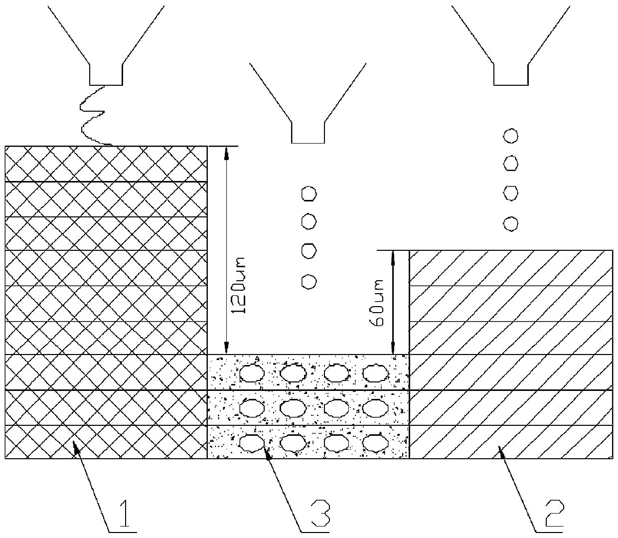
[0005] Another object of the present invention is to address the deficiencies in the prior art, to provide a combination of hard and soft natural polymer materials, and to use the differences in physical and chemical properties, curing mechanisms and time of different materials to establish multi-material composite molding. The multi-layer printing process solves the problem of mutual interference of materials in the complex structure forming process, and combines high bionic structure modeling and multi-degree-of-freedom robotic arm path planning to realize the 3D printing bionic modeling of cartilage-bone-bone marrow complex structures

Method used


Image

  • Cartilage-bone-marrow composite tissue structure based on 3D living cell printing and method of cartilage-bone-marrow composite tissue structure
  • Cartilage-bone-marrow composite tissue structure based on 3D living cell printing and method of cartilage-bone-marrow composite tissue structure
  • Cartilage-bone-marrow composite tissue structure based on 3D living cell printing and method of cartilage-bone-marrow composite tissue structure

Examples


Embodiment Construction

[0038] This embodiment discloses a method for preparing cartilage-bone-bone marrow composite tissue based on biological 3D printing, including the following steps:

[0039] 1. Biological information collection and modeling:

[0040] 1) By optimizing the bone structure image acquisition method, using perfusion casting and omics three-dimensional cross-sectional reconstruction technology, the three-dimensional data of the internal and external structures of the epiphysis of the human tibial plateau and blood circulation tube network are collected in a personalized manner;

[0041] 2) Input the collected biological information into computer software, express the actual tissue appearance and microenvironment as a bionic multi-material, multi-scale geometric model, and establish a bionic three-dimensional mathematical model of cartilage-bone-bone marrow and its microvessels.

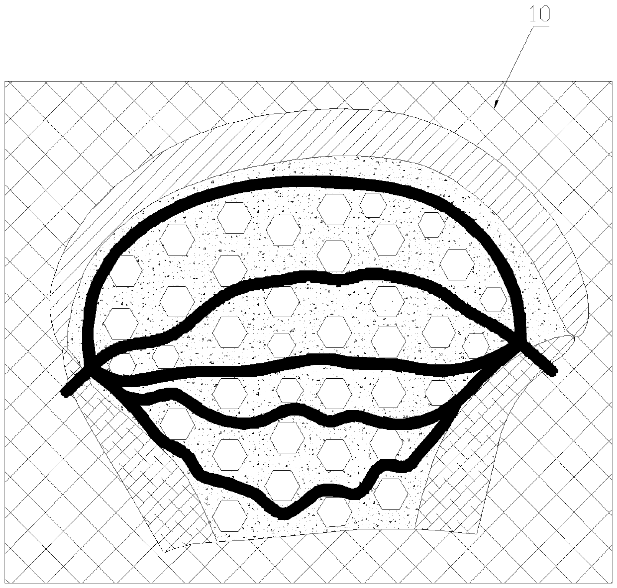
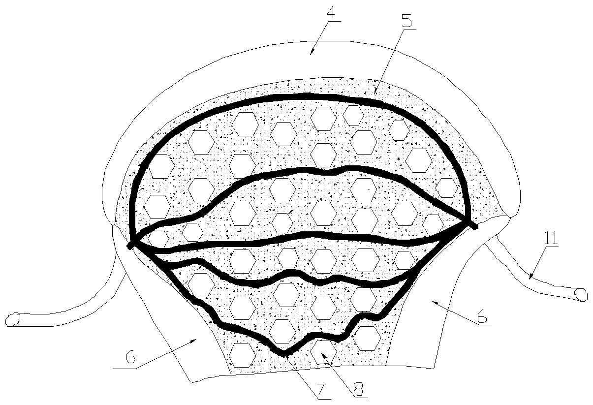
[0042] see figure 1 , figure 2 , Figure 4 , the geometric model includes:

[0043] a. Cartilage area...


PUM


Abstract

The invention discloses a cartilage-bone-marrow composite tissue structure based on 3D living cell printing and a method of the cartilage-bone-marrow composite tissue structure. Biological informationof bone tissue is collected and input into a computer for biomimetic modeling, and bio-ink suitable for cells such as cartilage, osteogenesis, mesenchymal stem, vascular endothelium, etc., is prepared; hard materials for printing dense bone area are prepared; various casting agent is prepared; living cells are inoculated into corresponding bio-ink at a certain density; and various types of printing ink are loaded into a printer bin correspondingly, 3D printing is carried out after setting printing parameters, and printed bone tissue is cultured in a tissue engineering bionic incubator. Aftera certain time, the method of temperature control or enzyme control or light control is used to remove casting agent materials at different times, artificial blood vessels are grafted on the outside of the cartilage-bone-marrow composite tissue, and continuous circulation perfusion cultivation is carried out in the tissue engineering biomimetic incubator, so as to achieve the pre-maturation of printed tissue, and the long-term survival and biological function of 3D printing bone tissue can be realized.

Description

technical field [0001] The invention belongs to biomedical engineering, in particular to a cartilage-bone-bone marrow composite tissue structure and method based on living cell 3D printing. Background technique [0002] Due to the high incidence of bone defects caused by war trauma and disease, a large number of bone graft prostheses are required clinically, and bone tissue transplantation ranks second among all tissue and organ transplants. At present, bone graft prostheses are mostly composed of metals and high molecular polymers, which only mimic the shape and appearance of defective bones, but have no biological activity, which often leads to serious defects in physiological functions after transplantation. In recent years, the rapid development of 3D printing technology provides the possibility to solve the above problems. Bio-3D printing is a promising method for constructing engineered human tissue, which can theoretically achieve a high degree of biomimetic tissue s...

Claims


Application Information

Patent Timeline
IPC IPC(8): A61L27/40A61L27/38A61L27/44A61L27/52A61L27/58B29C64/112B33Y10/00B33Y70/00B33Y80/00
CPCA61L27/3886A61L27/44A61L27/52A61L27/58A61L2430/02A61L2430/06B33Y10/00B33Y70/00B33Y80/00B29C64/112
Inventor 周强甘翼搏叶吉星李培涂兵罗磊赵晨欧阳斌张泽桐
Owner THE FIRST AFFILIATED HOSPITAL OF ARMY MEDICAL UNIV
Learn More