Self-assembly probe based on configuration change and unlabeled detection method for exosomes thereof

A technology for configuration change and detection method, applied in the field of molecular biology, can solve problems such as background interference and affecting the efficiency of identification

Active Publication Date: 2019-11-19
ZHENGZHOU UNIV
View PDF4 Cites 3 Cited by
  • Summary
  • Abstract
  • Description
  • Claims
  • Application Information

AI Technical Summary

Problems solved by technology

[0005] Patent CN201811548979.0 discloses an exosome detection method based on aptamer and rolling circle amplification, using an electrochemical sensor method to construct an aptamer probe based on exosome specificity, using G-quadruple Body-Hemin Mimetic Peroxidase Catalyzed H 2 o 2 The reaction generates signals; the patent synthesizes a large number of G-quadruplexes through rolling circle amplification for signal amplification to realize the quantitative detection of exosomes; patent CN201811173676.5 provides an aptamer group for detecting exosomes, lateral flow aptamer biosensor and its preparation method. The aptamer group for detecting exosomes in this patent includes CD63 aptamers and EpCam aptamers. CD63 aptamers can specifically bind to the CD63 protein of exosomes. EpCam aptamers The ligand can specifically bind to the EPCAM protein of exosomes. The invention also provides a lateral flow aptamer biosensor, which mainly sprays the CD63 aptamer on the binding pad based on the principle of chromatography test strips, and sprays the EpCam aptamer The detection line is formed, and the quality control line is sprayed with DNA probes that can be combined with nano-gold-labeled aptamers through base complementary pairing; the qualitative and quantitative detection of exosomes is realized through the lateral flow aptamer biosensor; now Some detections of exosomes are mostly based on the direct combination of antibodies or nucleic acid aptamers to exosomes surface general protein CD63, which will generate high background interference, and most of them need to label signal molecules , which will undoubtedly affect the efficiency of recognition, so it is imminent to develop an activatable, label-free fluorescent sensing platform to detect exosomes

Method used


Image

  • Self-assembly probe based on configuration change and unlabeled detection method for exosomes thereof
  • Self-assembly probe based on configuration change and unlabeled detection method for exosomes thereof
  • Self-assembly probe based on configuration change and unlabeled detection method for exosomes thereof

Examples


Embodiment 1

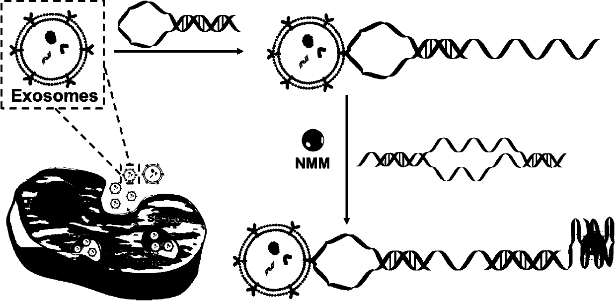
[0036] In this example, a self-assembly probe based on configuration change is used for a label-free detection method of exosomes, and the steps are as follows:

[0037] (1) Cell culture: CCRF-CEM, Ramos, Hela, B16F1 and HL-7702 cells were cultured in RPMI 1640 medium supplemented with 10% exosomes (fetal bovine serum removed) and 100IU / mL penicillin or streptomycin cultured at 37 °C with 5% wt / vol CO 2 Incubate in a humid incubator for 48 hours, and collect the cell supernatant when the cells grow to 80-90%;

[0038] (2) Isolation of exosomes: The cell supernatant obtained in step (1) was centrifuged at 3,000g for 30 minutes at 4°C, and filtered through a syringe-driven filter unit to remove intact cells and cell debris, collected by filtration and concentrated using 100KDaMWCO at 5,000g at 4°C for 30 minutes, then the filtered medium was ultracentrifuged at 160,000g for 2 hours at 4°C, the supernatant was discarded, and the exosome pellet was resuspended in phosphate buffered...

Embodiment 2

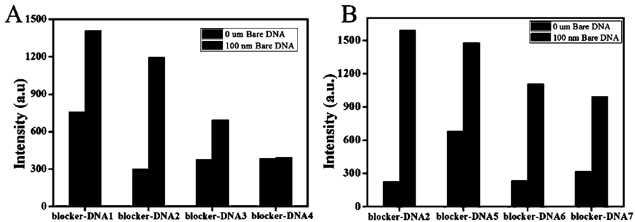
[0042] In this embodiment, in order to further study the hybridization and binding ability of G4-DNA and different blocker-DNAs, eight sets of Blocker DNA sequences are designed such as SEQ ID NO.3, SEQ ID NO.4, SEQ ID NO.5, SEQ ID NO .6, shown in SEQ ID NO.7, SEQ ID NO.8, SEQ ID NO.9 and SEQ ID NO.10; refer to the steps of Example 1 for fluorescence detection. First, a Bare DNA strand is designed, which contains the complete priming region bases, to simulate the probe recognition of exosomes, which can be used to trigger the strand displacement reaction and release G4. Test results such as figure 2 shown by figure 2 It can be seen that by using BlockerDNA-2, when there is Bare DNA, the maximum fluorescence enhancement can be obtained, which is significantly higher than that of blocker DNA-1, blockerDNA-3, blocker DNA-4, Blocker DNA-5, Blocker DNA-6 and Blocker DNA- 7. Therefore, blocker DNA-2 was selected as the best blocker DNA for subsequent experiments, and it was nam...

Embodiment 3

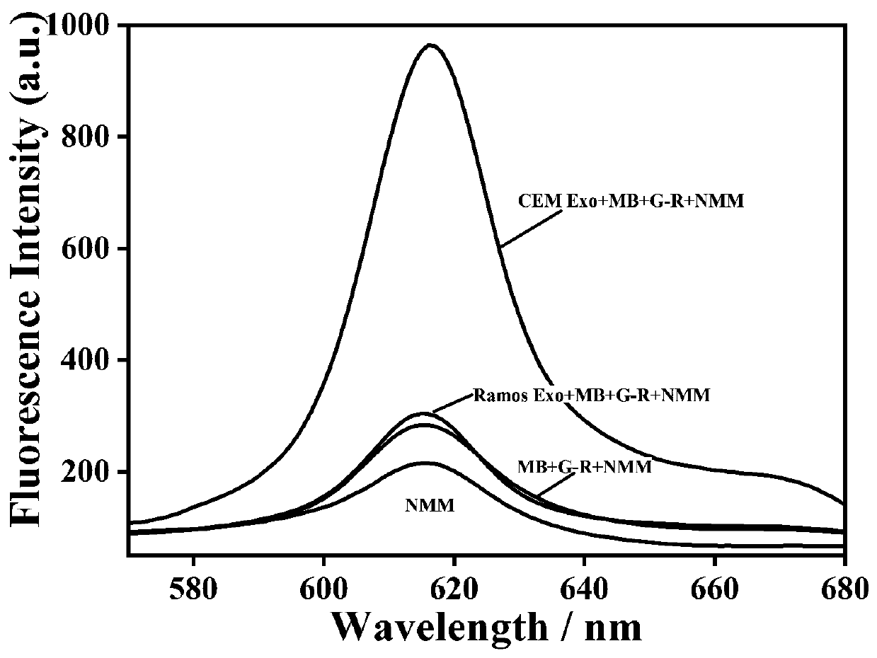
[0044] In this example, in order to prove the feasibility of the experimental principle, the fluorescence intensity of the system under different conditions was studied. The test results are shown in image 3 shown by image 3 It can be seen that in the absence of target exosomes (red curve) and control exosomes (blue curve), there is no obvious fluorescence signal, indicating that the present invention has a lower fluorescence background. After the target exosomes were introduced into the above mixed solution, a significant fluorescence enhancement could be observed around 615 nm (green curve), which indicated that the G-rich sequence was exposed and folded into a quadruple structure. These results demonstrate that this experimental platform can be used for the specific detection of leukemia-associated exosomes.


PUM


Abstract

The invention relates to the technical field of molecular biology, and in particular relates to a self-assembly probe based on configuration change and a label-free detection method for exosomes. Whenexosomes exist, are exosomes surface markers, protein tyrosine kinases-7 (PTK 7) specifically binds to the aptamer, changes the configuration of the recognition probe, releases the trigger chain, andthus binds to G-R DNA undergoes strand displacement reaction, exposing G-rich sequence to form G-quadruplex. NMM is embedded in G-quadruplex, generating strong fluorescence signal, realizing label-free sensitive detection of exosomes. Under the optimized experimental conditions, the linear range is 5*10<5> to 5*10<7> particles / mu L, and the minimum detection limit is 3.4*10<5> particles / mu L. Theprobe is used to prepare a highly sensitive and highly selective detection kit for detecting exosomes in actual blood samples.

Description

technical field [0001] The invention relates to the technical field of molecular biology, in particular to a self-assembly probe based on configuration change and a label-free detection method for exosomes. Background technique [0002] Exosomes are membrane-bound nanovesicles with diameters between 30 nm and 150 nm. They carry a large number of proteins, lipids, DNA, RNA and other biomolecules, making exosomes can be used as a non-invasive cancer marker for the preparation of early diagnosis reagents for tumors. Therefore, accurate quantification and classification of tumor exosomes is of great significance for the preparation of cancer diagnosis and prognosis assessment reagents. [0003] The performance analysis of different existing methods for detecting exosomes is summarized in the table below: [0004] . [0005] Patent CN201811548979.0 discloses an exosome detection method based on aptamer and rolling circle amplification, using an electrochemical sensor method ...

Claims


Application Information

Patent Timeline
IPC IPC(8): C12Q1/6876C12Q1/682C12N15/11
CPCC12Q1/6876C12Q1/682C12Q2531/119C12Q2563/107C12Q2525/205
Inventor 孟红敏陈娟李朝辉葛佳
Owner ZHENGZHOU UNIV
Why Eureka
Try Eureka