3D printing konjac glue gel stent, preparing method and application

A 3D printing, konjac gum technology, applied in medical science, prosthesis, additive processing, etc., can solve the lack of shear thinning properties of hydrogels, does not meet the needs of extrusion printing, and the stent is difficult to maintain the desired shape and other problems, to achieve the effect of controllable shape and microstructure of the stent, to achieve shape fidelity, and to achieve fidelity

Inactive Publication Date: 2019-11-22
GUANGXI MEDICAL UNIVERSITY
View PDF6 Cites 4 Cited by
  • Summary
  • Abstract
  • Description
  • Claims
  • Application Information

AI Technical Summary

Problems solved by technology

However, the application of these inks is limited. First, their hydrogels lack shear thinning properties. When printing, the printing pressure is high, which causes the composite cells to be under too high pressure, which affects the activity of cells, and then affects the regeneration and repair of tissues.
In addition, the precursor solutions of these inks exhibit low viscosity, which makes it

Method used


Image

  • 3D printing konjac glue gel stent, preparing method and application
  • 3D printing konjac glue gel stent, preparing method and application
  • 3D printing konjac glue gel stent, preparing method and application

Examples


example 1

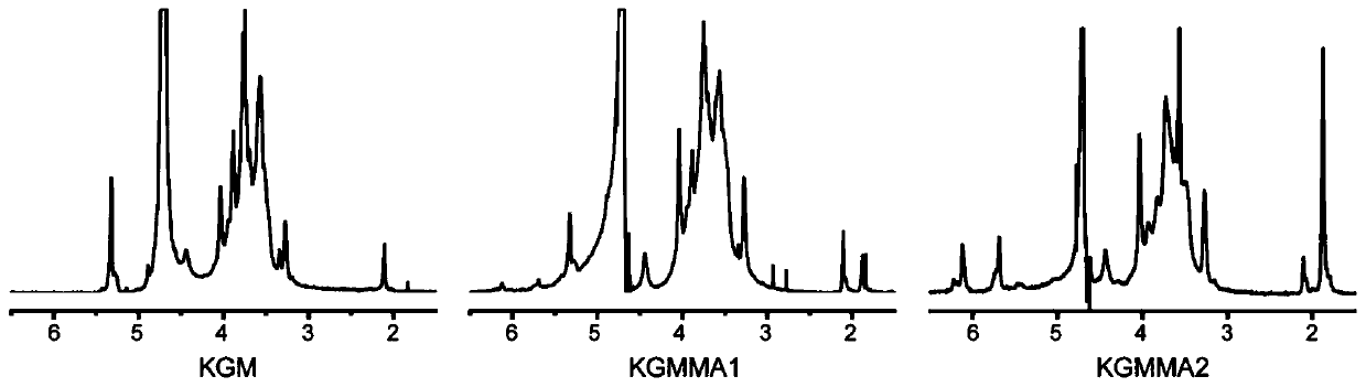
[0025] Wherein in the methacrylic anhydride modified konjac gum experiment, dropwise add 5M sodium hydroxide to maintain a certain pH to react, the pH selection is 7-8,8-9, prepare methacrylated konjac gum as follows: At 50°C, prepare 2% (w / v) konjac gum solution, add methacrylic anhydride, stir and mix evenly, then add 5M sodium hydroxide drop by drop to maintain pH 7-8, 8-9 respectively. After continuing to react for 6 hours, dialyze with ultrapure water for one week at 4°C and freeze-dry to obtain the product.

[0026] By comparing the degree of grafting of the product methacrylic anhydride, it was found that when the pH was 7-8, the efficiency of the grafted methacrylic anhydride of the product was higher.

Embodiment 2

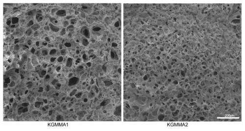
[0028] Wherein the printing concentration of konjac gum can be 6%, 7%, 8%, and the hydrogel of three kinds of concentrations prepares 3D printing support as follows: Use photoinitiator (0.1 %w / v) of PBS was completely dissolved to prepare a hydrogel; then the resulting hydrogel or chondrocytes (1*10 7 cell / ml) compound is extruded and printed through 3D-Bioplottersystem, and the ejected fiber filaments are piled up layer by layer to form a scaffold, and the printed scaffold is cross-linked by ultraviolet light for 60s to obtain the product.

[0029] Comparing the three scaffolds with printing parameters, it was found that the number of printed layers was between 8-10 layers, and 6% (w / v) required a lower printing voltage, which had less damage to the cell activity of composite cell printing .

example 3

[0031] The concentration of konjac gum raw material can be 1%-2% (w / v) when modified, and its preparation method is as follows: a kind of 3D printing konjac glue hydrogel support, and this support is prepared by the following method: konjac gum raw material is 50 Prepare a 1%-2% (w / v) aqueous solution of konjac gum at ℃, add different concentrations of methacrylic anhydride to modify it to prepare derivatives with different grafting degrees, dialyze the obtained product, and freeze-dry to obtain modified konjac gum.

[0032] By comparison, it was found that the degree of grafting of methacrylic anhydride in the solution with 2% concentration was higher than other concentrations.


PUM


Abstract

The invention discloses a 3D printing konjac glue gel stent, a preparing method and application. Konjac glue gel is methacrylated konjac glue gel. The preparing method includes the following steps ofmodifying a konjac glue gel raw material through methacrylic anhydride, conducting dialysis and freeze-drying to obtain methacrylated konjac glue gel powder, dissolving the methacrylated konjac glue gel powder in PBS with a photoinitiator to obtain hydrogel, combining the obtained hydrogel with chondrocyte for printing through a 3D-Bioplotter system, and then conducting ultraviolet crosslinking toobtain the konjac glue gel stent product. The stent combined with the chondrocyte has the potential for cartilage repair engineering. The 3D printing konjac glue gel stent has the effects of being good in biocompatibility, controllable in material performance, low in cost, wide in source and simple in operation.

Description

technical field [0001] The invention relates to the technical field of biocomposite materials, in particular to a 3D printed konjac glue hydrogel scaffold and its preparation method and application. Background technique [0002] Tissue engineering provides a new treatment method for the regeneration and repair of human tissues and organs. Tissue engineering technology includes electrospinning and 3D printing technology. The former mainly provides a two-dimensional microenvironment for cells, which is far from satisfying the requirements of bionic tissue. The 3-dimensional microenvironment of the organ, while the latter can be achieved. 3D bioprinting technology can accumulate biomaterials with micron-level precision, regulate the spatial distribution of living cells and functional molecules, and then reshape the complex physiological microenvironment of 3D tissues and organs. However, the lack of individualized biochemical and mechanical properties of biomaterials is still ...

Claims


Application Information

Patent Timeline
IPC IPC(8): A61L27/20A61L27/38A61L27/52C08F251/00C08F220/08B33Y70/00
CPCA61L27/20A61L27/3817A61L27/3852A61L27/52A61L2430/06B33Y70/00C08F251/00C08F220/08C08L51/02
Inventor 郑立赵劲民覃再嫩庞运芬梁锐明黄权新
Owner GUANGXI MEDICAL UNIVERSITY
Why Eureka
Try Eureka