Preparation and application of an ultra-bright fluorescent carbon dot
A fluorescent carbon dot and ultra-bright technology, applied in the field of nanomaterials and biology, can solve the problems of low fluorescence quantum yield, high synthesis cost, and short imaging time, and achieve the effect of simple operation method, low cost, and simple operation
- Summary
- Abstract
- Description
- Claims
- Application Information
AI Technical Summary
Problems solved by technology
Method used
Image
Examples
preparation example Construction
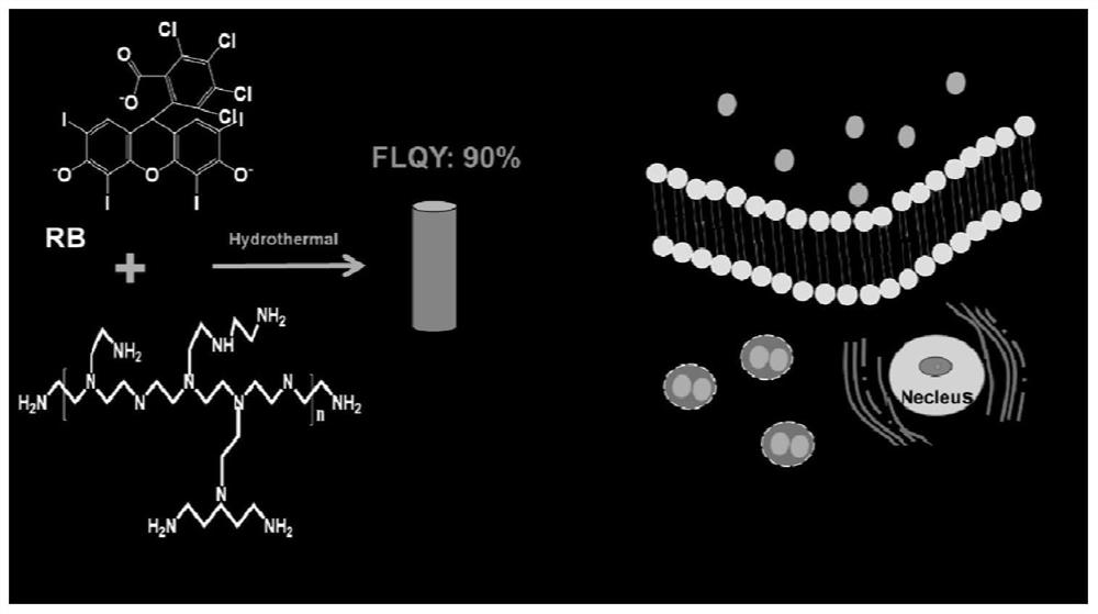
[0047] The preparation method of the above-mentioned ultra-bright fluorescent carbon dots comprises the following steps:
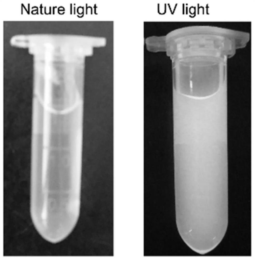
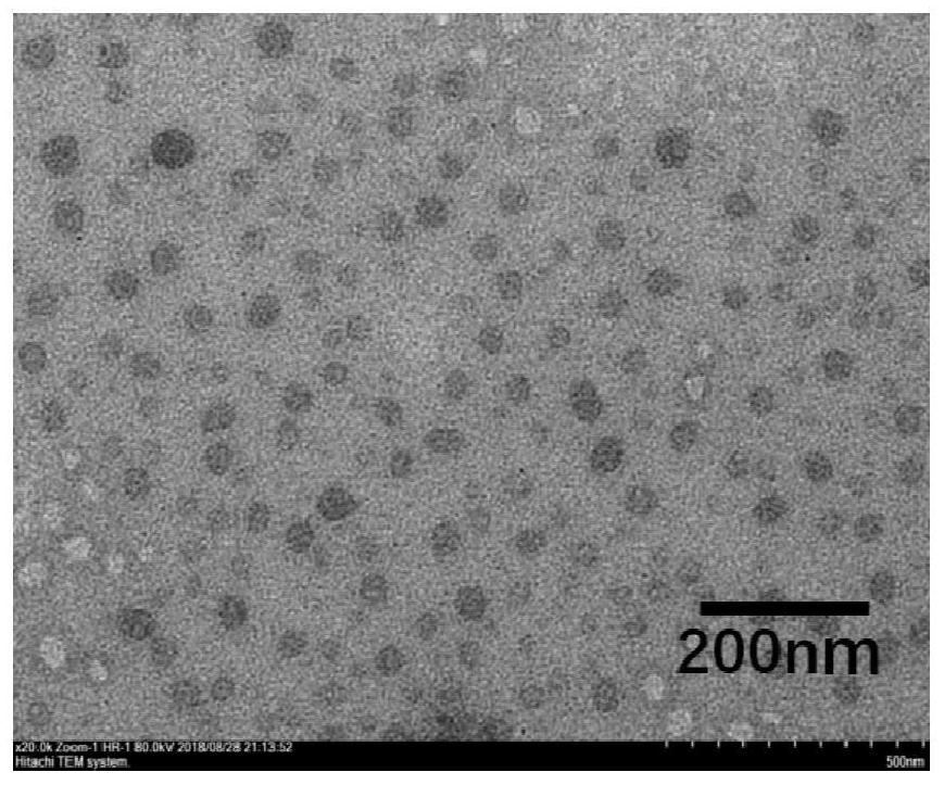
[0048] Dissolve the formula amount of RB in ultrapure water, add the formula amount of bPEI to react; after the reaction is completed, the reaction solution cooled to room temperature is dialyzed to obtain high-purity fluorescent carbon dots.
[0049] in,
[0050] The reaction temperature is 25-200°C, preferably 160°C;
[0051] The reaction time is 2~24h, preferably 6h;
[0052] Raw material concentration ratio (bPEI:RB) 0.5:1~5:1, preferably 3:1
[0053] The reaction device is preferably a hydrothermal reactor.
[0054] Wherein the molecular weight cut-off of the dialysis bag used for dialysis is 500-1000.
[0055] The application of the above-mentioned ultra-bright fluorescent carbon dots as lysosome-targeted fluorescent probes also falls within the protection scope of the present invention.
Embodiment 1
[0058] Synthesis of P-R CDs:
[0059] 1 mL of bPEI diluent was mixed with 4 mL of 15 mg / mL RB solution, and the mixture was transferred to a 10 mL autoclave, hydrothermally reacted at 160 °C for 6 h, cooled to room temperature, and the crude P-RCDs solution was obtained by intercepting Dialysis with a dialysis belt with a molecular weight of 500-1000 for 12 hours.
Embodiment 2
[0061] Colocalization analysis of P-R CDs for lysosome-targeted imaging:
[0062] In an incubator, HL-7702 cells were incubated with 150 μL, 1 mg / mL of P-R CDs for 10, 20, 30, and 60 minutes, respectively. When the incubation was completed, the old medium was discarded, washed three times with PBS, and fresh medium was added for imaging. . In particular, before imaging, commercial Lyso Tracker Red (Lyso Red) dye needs to be added half an hour in advance to visualize lysosomes, and SP8 confocal microscope is used for fluorescence imaging analysis.
PUM
 Abstract
Description
Claims
Application Information
 - R&D
- Intellectual Property
- Life Sciences
- Materials
- Tech Scout
- Unparalleled Data Quality
- Higher Quality Content
- 60% Fewer Hallucinations
Browse by: Latest US Patents, China's latest patents, Technical Efficacy Thesaurus, Application Domain, Technology Topic, Popular Technical Reports.
© 2025 PatSnap. All rights reserved.Legal|Privacy policy|Modern Slavery Act Transparency Statement|Sitemap|About US| Contact US: help@patsnap.com


