Dissociation and separation method for shellfish spermary spermatogenic cells

A technology for spermatogenic cells and shellfish, which is applied in the field of separation of marine shellfish cells, can solve the problems of difficult separation of shellfish spermatogenic cells, and achieves the effects of large application value, high content and improved activity.

Active Publication Date: 2020-08-14
OCEAN UNIV OF CHINA
View PDF6 Cites 2 Cited by
  • Summary
  • Abstract
  • Description
  • Claims
  • Application Information

AI Technical Summary

Problems solved by technology

[0004] In view of this, the purpose of the present invention is to provide a method for dissociation and separation of shellfish testicu

Method used


Image

  • Dissociation and separation method for shellfish spermary spermatogenic cells
  • Dissociation and separation method for shellfish spermary spermatogenic cells
  • Dissociation and separation method for shellfish spermary spermatogenic cells

Examples


Embodiment 1

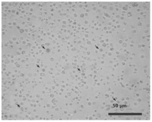
[0047] Take the testes of Chlamys farreri in the proliferating stage, and cut the testes into 1mm after cleaning and disinfection 3 The small pieces were then digested with 0.1% collagenase IV, and then digested with 0.25% trypsin. The time of the two enzymes for digestion is shown in Table 1. The testis was dissociated to obtain a single-cell suspension of Chlamys farreri germ cells.

[0048] The results are shown in Table 1. The results showed that at 25°C, when the concentrations of the two enzymes (0.1% collagenase IV and 0.25% trypsin) were fixed, the number of cells obtained by dissociation of 0.1 g of testis gradually increased with the prolongation of the treatment time of the two enzymes. increased, but the viability of cells gradually decreased after dissociation. Among them, experimental group 1 (digested with 0.1% collagenase IV for 60 min and digested with 0.25% trypsin for 5 min) had the highest survival rate of cells after dissociation, which was 98.23%, but th...

Embodiment 2

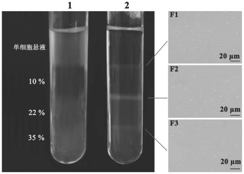
[0054] 1. Obtaining single cells from the testis of Chlamys farreri

[0055] ① Treatment of testicular tissue

[0056] Select the proliferating male clamshell scallop with good vitality and slightly white testes, wipe the surface of the double shell with 75% alcohol to disinfect, and then dissect out the testis tissue in an ultra-clean workbench, use calcium and magnesium ion-free phosphate buffer for cells (1 ×CPBS) rinsed 3 times, transferred to citrate-modified phosphate buffer solution (1×CPBS solution) containing double antibodies (350IU / ml penicillin and 350IU / ml streptomycin) and soaked for 1 hour for sterilization and disinfection, and then rinsed with 1×CPBS solution 3 times, use ophthalmic scissors to cut the testis tissue into a volume of 1mm 3 Little pieces left and right.

[0057] ② Preparation of single cell suspension by two-step enzymatic hydrolysis

[0058] Collect 0.1g of the shredded testis tissue into a 2ml EP tube, add 1ml of 0.1% collagenase IV, digest...


PUM

PropertyMeasurementUnit
Diameteraaaaaaaaaa
Cell diameteraaaaaaaaaa

Abstract

The invention provides a dissociation and separation method for shellfish spermary spermatogenic cells, and belongs to the technical field of the separation of ocean shellfish cells. The dissociationand separation method comprises the following steps of: the spermary of a shellfish during a proliferating phase is broken and is digested by a collagenase IV solution of which the mass concentrationis 0.1% and a trypsin solution of which the mass concentration is 0.25% in sequence, and spermary dissociation is carried out to obtain a single-cell suspension; and Percoll solutions of which the volume concentration is independently 10%, 22% and 35% is prepared, and the Percoll solutions are loaded in the same centrifuge tube in a descending order, the single-cell suspension of the shellfish spermatogenic cells is added into the top layer of the centrifuge tube, and centrifugation is carried out to independently collect three layers of cells. By use of a two-step enzymolysis method, spermatogenic cells in spermary tissues can be successfully dissociated into the single-cell suspension, a cell survival rate can be as high as 96.62%, and the cells can be normally subjected to in vitro normal culture. Meanwhile, high-purity spermatogonia and spermatocytes can be separated, an important material is provided for researching a shellfish gamete generating and breeding mechanism, and the dissociation and separation method has a high guidance meaning and a good application value.

Description

technical field [0001] The invention belongs to the technical field of separation of marine shellfish cells, and in particular relates to a method for dissociation and separation of shellfish testicular germ cells. Background technique [0002] Sperm cells are collectively referred to as male germ cells, which can form spermatozoa through graded differentiation, transmit genetic information to offspring, and maintain species stability. According to the process of development and differentiation, germ cells can be divided into spermatogonia, primary spermatocytes, secondary spermatocytes, sperm cells and spermatozoa. The study of germ cells has always been a hot spot in developmental biology and reproductive biology. The in vitro isolation and purification of germ cells is conducive to exploring the mechanism of spermatogenesis and realizing the in vitro culture and transplantation of germ cells. However, due to the small size and difficulty of identifying germ cells, it is ...

Claims


Application Information

Patent Timeline
IPC IPC(8): C12N5/07
CPCC12N5/0601C12N2509/00
Inventor 秦贞奎王庆张志峰刘丹雯
Owner OCEAN UNIV OF CHINA
Why Eureka
Try Eureka