High-flux integrated phosphorylated proteomics detection method

A phosphorylation and analysis method technology, applied in the field of proteomics, can solve the problems of limited application, large amount of initial protein and other materials, etc., to achieve the effect of improving throughput and reducing sample analysis time

Inactive Publication Date: 2020-08-25
SOUTH UNIVERSITY OF SCIENCE AND TECHNOLOGY OF CHINA
View PDF5 Cites 2 Cited by
  • Summary
  • Abstract
  • Description
  • Claims
  • Application Information

AI Technical Summary

Problems solved by technology

However, these methods require large amounts of starting protein and other materials, which limits their application to large-scale samples

Method used


Image

  • High-flux integrated phosphorylated proteomics detection method
  • High-flux integrated phosphorylated proteomics detection method
  • High-flux integrated phosphorylated proteomics detection method

Examples


Embodiment 1

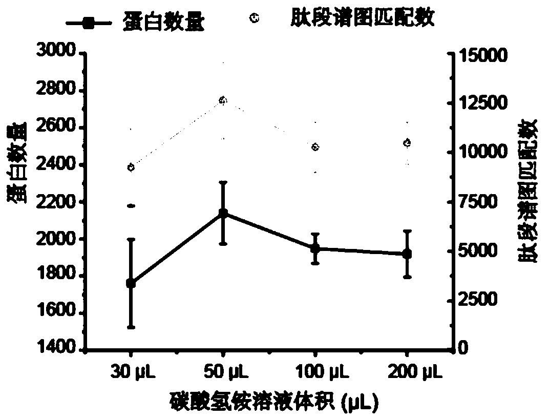
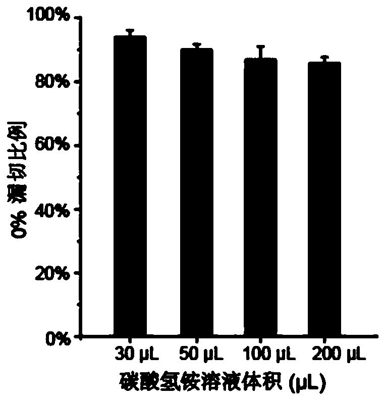
[0050] The optimization of different ammonium bicarbonate solution volumes to enzymolysis efficiency of embodiment 1

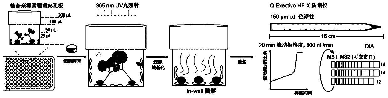
[0051] This example aims to realize the capture and analysis of related tyrosine phosphorylated proteins in a small amount of cell lysate, reduce the concentration of cell lysate, and test the feasibility and sensitivity of the method. The overall operation process is as follows figure 1 .

[0052] Add the corresponding amount of probe to the bait protein SH2 domain (1 mg / mL, dissolved in HEPES buffer) at a molar ratio of 1:10 (protein:probe), and incubate at room temperature for 2 minutes. Subsequently, 1 mol / L glycine solution was added according to the molar ratio of 1:10 (probe: glycine) to terminate the reaction between the probe and the bait protein. The resulting probe-labeled bait protein was ultrafiltered 7 times to remove unreacted probe and glycine molecules. Determine the protein concentration and dilute to 0.02 μg / μL. The bait protein labeled ...

Embodiment 2

[0054] Example 2 Exploration of the sensitivity of different cell dosages to sample pretreatment methods

[0055] According to the method in Example 1, 5 μg, 50 μg, and 300 μg of HeLa cell lysate before and after stimulation with EGF were added to the streptavidin-coated 96-well plate to which the probe and bait protein were connected (including 3 technical repetitions). ), and then carry out subsequent processing according to the general conditions described in Example 1, and carry out the comparison of protein complex enrichment and identification under EGF stimulation conditions.

[0056] The result is as Figure 3A As shown, as the amount of cell lysate increases, more protein complexes are enriched and identified after EGF stimulation. Among them, 17 EGFR signaling pathway-related proteins were enriched in only 5 μg of cell lysate. When the amount of cell lysate increased to 50 μg and 300 μg, 68 and 92 stimulation-related proteins could be enriched after EGF stimulation,...

Embodiment 3

[0058] Embodiment 3 method comparison

[0059] According to the method determined in Example 1 and Example 2, add 50 μg of HeLa cell lysate before and after EGF stimulation to the 96-well plate coated with streptavidin that has been connected to the probe and bait protein (including 3 times of technology) Repeat), then carry out follow-up treatment by general condition described in embodiment 1. On the other hand, in a 1.5mL centrifuge tube, add the same mass of bait protein linked to the probe, according to the patent 201910599017.6 treatment method, add 50μg of HeLa cell lysate before and after EGF stimulation (including 3 technical repetitions), and use 10μL streptavidin Avidin beads were used for capture, followed by subsequent processing according to the general conditions described in the patent 201910599017.6. The samples treated by the two methods were compared for the enrichment and identification of protein complexes under EGF stimulation conditions.

[0060] The r...


PUM

PropertyMeasurementUnit
concentrationaaaaaaaaaa

Abstract

The invention provides a high-flux integrated phosphorylated proteomics detection method. A capture skeleton is formed by a probe connected to a streptavidin plate and bait protein, a tyrosine phosphorylated protein compound is enriched, captured and subjected to enzymolysis to obtain polypeptide, and the sensitivity is improved by 300 times under the condition that the use amount of the probe isreduced by 100 times. Short-gradient rapid separation is realized by utilizing high performance liquid chromatography of a self-filling chromatographic column with the inner diameter of 150 microns; efficient detection is carried out in a DIA data acquisition mode by combining a Q Extractive HF-X mass spectrometer, and separation and detection of a single sample within 35 minutes can be realized under the condition of not losing qualitative and quantitative detection performance. Identification of transient changes of phosphorylation conditions of related signal channel proteins in cells underthe action of inhibitors with different concentrations in different EGF stimulation time is realized. And a high-flux, high-sensitivity and rapid detection method is provided for enrichment and identification of the tyrosine phosphorylated protein compound.

Description

technical field [0001] The invention relates to the technical field of proteomics, to a high-throughput, integrated tyrosine phosphorylated protein capture method and its application, and in particular to a highly sensitive integrated sample pretreatment method and rapid mass spectrometry detection method. Background technique [0002] The abnormal activation of phosphorylated tyrosine (pTyr) protein and its mediated signaling protein complex often reveals the activation of disease-related signaling pathways, and such proteins are often used as biomarkers and drug targets. Therefore, the capture and detection of such protein complexes play an important role in the discovery of biomarkers and drug targets. [0003] The protein interaction research method combined with affinity purification and mass spectrometry is currently widely used to capture and identify such protein complexes. Usually, a key signaling protein with a specific tag is expressed in living cells, and the pr...

Claims


Application Information

Patent Timeline
Patent Type & Authority Applications(China)
IPC IPC(8): G01N33/68G01N30/02G01N30/72
CPCG01N30/02G01N30/72G01N33/6848
Inventor 田瑞军李鹏飞孔倩
Owner SOUTH UNIVERSITY OF SCIENCE AND TECHNOLOGY OF CHINA
Why Eureka
Try Eureka