Application of USP33 as drug target in preparation of drugs

A technology for preparing drugs and drugs, applied in the field of protein engineering, can solve problems such as reducing the survival rate of Drosophila

Active Publication Date: 2020-10-09
BEIJING INST OF GENOMICS CHINESE ACAD OF SCI CHINA NAT CENT FOR BIOINFORMATION
View PDF5 Cites 2 Cited by
  • Summary
  • Abstract
  • Description
  • Claims
  • Application Information

AI Technical Summary

Problems solved by technology

After infection with L.monocytogenes in Drosophila, the function of LC3 in Drosophila lacking PARK2 will be reduced, resulting in the loss of autophagy and reducing the survival rate of Drosophila

Method used


Image

  • Application of USP33 as drug target in preparation of drugs
  • Application of USP33 as drug target in preparation of drugs
  • Application of USP33 as drug target in preparation of drugs

Examples


Embodiment 1

[0096] Example 1 Co-immunoprecipitation and mass spectrometry identification of the interaction between Parkin and the deubiquitinating enzyme USP33

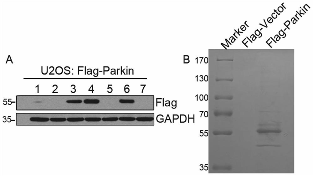
[0097] 1. The present invention utilizes co-immunoprecipitation combined with mass spectrometry to study Parkin's interacting protein network. In U2OS cells, the pcDNA3-Flag-Parkin plasmid was transfected, and the cells were screened by 400ug / ml G418 to obtain a cell line stably expressing the Flag-Parkin protein SEQ ID NO.2 ( figure 1 Figure A).

[0098] Cell No. 4 with a high expression level was selected for expansion culture, the whole cell lysate was collected, and anti-FlagBeads were incubated with it, the Flag-Parkin interacting protein was enriched by co-immunoprecipitation, and SDS-PAGE gel electrophoresis was used Separation of enriched protein samples ( figure 1 Figure B), and then stained with Coomassie Brilliant Blue G250 for protein profile analysis. The control group was transfected with pcDNA3-Flag-vector plas...

Embodiment 2

[0105] Example 2 Mitochondrial localization of USP33 protein

[0106] 1. USP33 is located in the mitochondrial outer membrane

[0107] The localization of endogenous USP33 in cells was verified by immunofluorescence experiments. In U2OS cells, mitochondria were labeled with Tom20 (mitochondrial outer membrane protein) antibody, and U2OS cells were labeled with USP33 antibody, as Figure 5 As shown in Figure A, it was found that endogenous USP33 and mitochondrial protein TOM20 had obvious co-localization, indicating that USP33 protein was located on mitochondria. Next, cell fractionation was performed on HEK293 cells, as Figure 5As shown in Figure B, the contamination of nuclear LaminB and cytoplasmic Tubulin was not detected in the mitochondrial fraction, indicating that the isolated mitochondria are of good purity and can be used in the next experiment. At the same time, Western blotting experiments confirmed that USP33 exists in mitochondria, as shown in the immunofluores...

Embodiment 3

[0111] Example 3 Knockdown of USP33 Promotes GFP-Parkin Translocation to Mitochondria

[0112] When cells are treated with CCCP (Carbonyl cyanide 3-chlorophenylhydrazone, inhibitor of mitochondrial oxidative phosphorylation), the mitochondria will be damaged and depolarized, and a large amount of Parkin protein will be recruited to the mitochondria to initiate the occurrence of mitophagy. In order to study whether the knockdown of USP33 will affect the process of mitophagy initiation, this example constructed a GFP-Parkin plasmid, transfected it into U2OS cells, and established a cell line stably expressing GFP-Parkin.

[0113] Cells stably expressing GFP-Parkin were treated with different concentrations of CCCP for 2 hours, and then the translocation of GFP-Parkin was observed, as shown in Figure 7 As shown, compared with the control group, GFP-Parkin aggregated and translocated in most of the cells treated at the concentration of 20 μM and 40 μM, while GFP-Parkin aggregated...


PUM


Abstract

The invention provides an application of USP33 as a medication target in preparation of drugs, and belongs to the field of protein engineering. According to the invention, the deubiquitination enzymeUSP33 interacting with Parkin is discovered by utilizing co-immunoprecipitation and mass spectrometry technologies, so that the USP33 is further discovered to be positioned on the outer membrane of mitochondria, and the E3 ligase Parkin can be modified by deubiquitination. When the USP33 is knocked down, Parkin transposition to mitochondria can be promoted; therefore, mitochondrial autophagy is enhanced to remove damaged mitochondria; USP33 regulates mitochondrial autophagy by influencing deubiquitination of Parkin, apoptosis of nerve cells under MPTP treatment can be inhibited by knocking down USP33, USP33 can be used as a medication target for preparing a medicine, and good application potential and value are achieved.

Description

technical field [0001] The invention belongs to the field of protein engineering, and specifically relates to the use of USP33 as a drug target in the preparation of medicines. Background technique [0002] USP33 (VDU1) is a deubiquitinating enzyme with 942 amino acids and a molecular size of 107kD. It can remove ubiquitin molecules from the substrate, and can also rely on pVHL (mutant protein of von Hippel–Lindau (VHL) disease) way to be ubiquitinated. Structural biology revealed that it contains a ZnF UBP structure at the N-terminus, a similar structure is also found in the deubiquitinating enzyme HDAC6, and a DUSP structure at the C-terminus. The ZnF UBP structure has three zinc ions, and the 221-arginine mutation of USP33 will affect the binding to the 75-glycine ubiquitin molecule. USP33 has at least three variants. Variant 1 is full-length and has 25 exons; variant 2 lacks exon 2, and translation starts from the start codon of exon 3 lacking 31 amino acids; Body 3 i...

Claims


Application Information

Patent Timeline
Patent Type & Authority Applications(China)
IPC IPC(8): C12N9/00G01N33/573A61K38/48A61K38/53A61K45/00A61P3/00A61P9/00A61P25/28A61P31/12A61P35/00
CPCC12N9/93G01N33/573C12Y603/02019A61K38/4813A61K38/53A61K45/00A61P25/28A61P35/00A61P9/00A61P3/00A61P31/12G01N2333/948G01N2500/10
Inventor 赵永良牛凯峰方洪波魏迪
Owner BEIJING INST OF GENOMICS CHINESE ACAD OF SCI CHINA NAT CENT FOR BIOINFORMATION
Why Eureka
Try Eureka