Preparation method and application of tissue autofluorescence quenching agent

A technology of autofluorescence and fluorescence quenching, applied in the preparation of test samples, fluorescence/phosphorescence, material excitation analysis, etc., which can solve the problems of long time and lack of equipment.

Inactive Publication Date: 2020-10-23
北京基谱生物科技有限公司
View PDF2 Cites 3 Cited by
  • Summary
  • Abstract
  • Description
  • Claims
  • Application Information

AI Technical Summary

Problems solved by technology

This method takes a long time, and most immunohistochemical laboratories do not have the equipment required for this method

Method used


Image

  • Preparation method and application of tissue autofluorescence quenching agent
  • Preparation method and application of tissue autofluorescence quenching agent
  • Preparation method and application of tissue autofluorescence quenching agent

Examples


Embodiment 1

[0034] Preparation of Tissue Autofluorescence Quencher A: Weigh 8.0g of NaCl, 0.2g of KCl, 1.44g of Na2HPO4, and 0.24g of KH2PO4, add ultrapure water to dissolve to a constant volume of 1000ml, and obtain a phosphate buffer solution with a pH of 7.4. Weigh 10 mg of sodium borohydride and dissolve in 10 ml of phosphate buffer solution with a pH of 7.4.

[0035] Preparation of Tissue Autofluorescence Quencher B: Weigh 0.5 g of Sudan Black B, dissolve in 70% alcohol for 2 days, filter for later use.

Embodiment 2

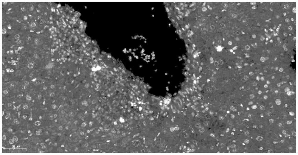
[0037] Method of use of tissue autofluorescence quencher: mouse liver CD8 immunofluorescence

[0038] S1. Paraffin sections were dewaxed to water: put the sections in xylene I for 15 minutes, xylene II for 15 minutes, absolute ethanol I for 5 minutes, absolute ethanol II for 5 minutes, 85% alcohol for 5 minutes, 75% alcohol for 5 minutes, and distilled water to wash;

[0039] S2. Antigen retrieval: place the tissue slices in a repair box filled with EDTA antigen retrieval buffer (pH 8.0) and carry out antigen retrieval in a steamer. When the temperature reaches 95°C, time 30 minutes. During this process, excessive evaporation of the buffer should be prevented. Do not dry the slides. After natural cooling, place the slides in PBS (pH 7.4), shake and wash 3 times on a decolorizing shaker, 5 minutes each time;

[0040] S3. Circle autofluorescence quenching: After the slices are dried slightly, draw a circle around the tissue with a histochemical pen to prevent the antibody from f...

Embodiment 3

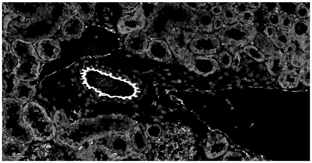
[0049] Method of use of tissue autofluorescence quencher: mouse kidney CD31 immunofluorescence

[0050] S1. Paraffin sections were dewaxed to water: put the sections in xylene I for 15 minutes, xylene II for 15 minutes, absolute ethanol I for 5 minutes, absolute ethanol II for 5 minutes, 85% alcohol for 5 minutes, 75% alcohol for 5 minutes, and distilled water to wash;

[0051]S2. Antigen retrieval: place the tissue slices in a repair box filled with EDTA antigen retrieval buffer (pH 8.0) and carry out antigen retrieval in a steamer. When the temperature reaches 95°C, time 30 minutes. During this process, excessive evaporation of the buffer should be prevented. Do not dry the slides. After natural cooling, place the slides in PBS (pH 7.4), shake and wash 3 times on a decolorizing shaker, 5 minutes each time;

[0052] S3. Quenching autofluorescence by drawing a circle: After the slices are dried slightly, draw a circle around the tissue with a histochemical pen to prevent the a...


PUM


Abstract

The invention discloses a tissue autofluorescence quenching agent. The tissue autofluorescence quenching agent is prepared from a tissue autofluorescence quenching agent A and a tissue autofluorescence quenching agent B, wherein the tissue autofluorescence quenching agent A is a mixture of a phosphoric acid buffer solution with the pH value of 7.4 and sodium borohydride; and the tissue autofluorescence quenching agent B is a mixed solution of Sudan black B and absolute ethyl alcohol. An application method of the tissue autofluorescence quenching agent comprises the following steps: S1, dewaxing paraffin sections to water: sequentially putting the sections into xylene I for 15 minutes, xylene II for 15 minutes, absolute ethyl alcohol I for 5 minutes, absolute ethyl alcohol II for 5 minutes,85% alcohol for 5 minutes and 75% alcohol for 5 minutes, and washing with distilled water; and S2, repairing antigen: placing the tissue sections in a repair box full of an EDTA antigen repair buffersolution. According to the preparation and the application of the tissue autofluorescence quenching agent, the preparation process of the agent is simple and rapid, the raw materials are easy to obtain, the tissue autofluorescence can be effectively reduced, and the signal-to-noise ratio in immunofluorescence analysis is improved.

Description

technical field [0001] The invention relates to the application field of biomedicine, in particular to the preparation and application of a tissue autofluorescence quencher. Background technique [0002] Immunofluorescence technology is an experimental technique based on the specific combination of antibodies and antigens. It is widely used in basic research and clinical experiments due to its advantages of simple operation, high efficiency, and strong specificity. However, the autofluorescence in the tissue causes great interference to the detection results of tissue sections, and even leads to the failure of staining. The main reason for tissue autofluorescence is from the components of the tissue itself. It mainly includes rich endogenous peroxidase, collagen, elastin, lipofuscin and so on. Apart from the tissue section itself, fixatives such as formaldehyde, glutaraldehyde, and formalin often used in tissue fixation can also cause tissue autofluorescence. [0003] One...

Claims


Application Information

Patent Timeline
Patent Type & Authority Applications(China)
IPC IPC(8): G01N1/30G01N1/28G01N21/64G01N33/533G01N33/58
CPCG01N1/30G01N1/28G01N21/6428G01N33/533G01N33/582G01N2001/302
Inventor 池喜峰来丽丽王宇赵成成
Owner 北京基谱生物科技有限公司
Why Eureka
Try Eureka