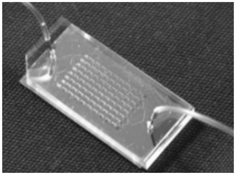
Preparation method of microfluid system for capturing and enriching circulating tumor cells and microfluid device
A microfluidic system and tumor cell technology, applied in the preparation of microfluidic systems and the field of microfluidic devices, can solve the problem of low sensitivity, meet the requirements of reducing interference and quantity, and realize all-round detection and high efficiency. the effect of capturing
- Summary
- Abstract
- Description
- Claims
- Application Information
AI Technical Summary
Problems solved by technology
Method used
Image
Examples
Embodiment 1
[0061] The present invention proposes a preparation method of a microfluidic system for capturing and enriching circulating tumor cells, which is prepared by the following preparation method: (this embodiment uses a 0.5 mm thick medical pathological quartz glass slide as the basis)
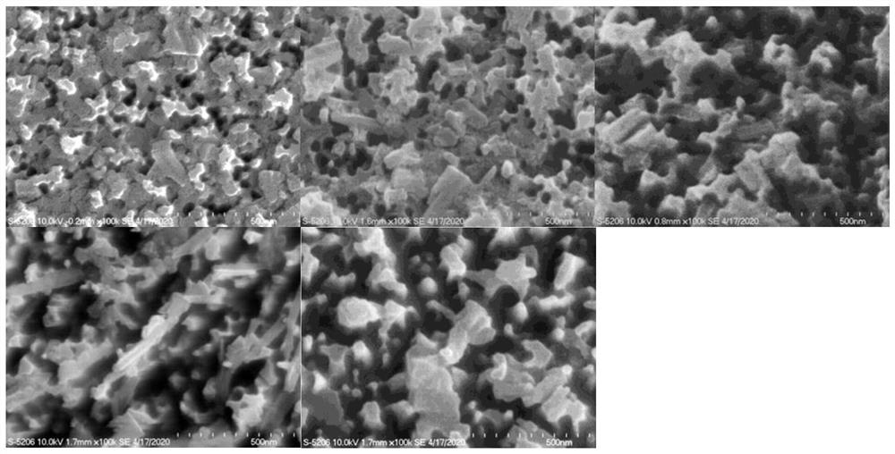
[0062] 1) Etching, surface modification and biofunctionalization of silicon nanowire chips
[0063] see Figure 1 to Figure 5 , is the etching process of the silicon nanowire chip, and the specific steps are as follows:
[0064] S1, spin coating PMMA-A8 photoresist glue on the pathological glass slide, the thickness of the PMMA-A8 photoresist glue is 500nm, dry for later use;
[0065] S2, carry out O to the PMMA-A8 photoresist glue of coating 2 Reactive ion etching to form point-like nanostructures, wherein the etching time is 2 minutes;
[0066] S3. Carry out CF4 etching to the dot-shaped nanostructure, etch for 10 minutes, and wash with concentrated sulfuric acid / hydrogen peroxide mixed solut...
Embodiment 2
[0087] 1) Preparation of simulated samples
[0088] Prepare a cell suspension with a density of 10,000 / mL (accurate count) from the culture dish of the non-small cell lung cancer cell line H2228 with a culture abundance of about 95%, and dilute and sort 200 / 20uL of the ready-to-use solution in a gradient, and add 180uL Contains 2*10 5 Jurkat cell suspension (the cells are all diluted and configured with PBS), as a 200uL mock sample. Add 1.5mL solution containing 5umol biotin-labeled EpCAM antibody (anti-EpCAM) to the mock sample, and incubate at room temperature for 45 minutes. Then centrifuge (400g, 5 minutes), after removing the supernatant, wash again with PBS, and constant volume in 200uL PBS solution, as test simulation sample.
[0089] 2) Test and selection of optimal flow rate
[0090] ①Experimental steps:
[0091] Select the simulated sample in experiment 1) as the test object, and test the capture efficiency of H2228 cells at flow rates of 0.1mL / h, 0.2mL / h, 0.5mL / h,...
Embodiment 3
[0111] 1) Testing and application of blood samples from clinical cancer patients:
[0112] ①The blood collection of clinical samples requires BD Vacutainer Glass ACD Solution A tube8.5mL to avoid EDTA anticoagulant damage to the antigen on the surface of the cells in the blood, which will affect the binding of the capture antibody and reduce the capture efficiency; take the first tube of blood draw only Take 2mL and not use it for the test (to detect false positive cells due to epithelial cells exfoliated when the needle is inserted into the blood).
[0113] ② Taking 4mL whole blood as an example, this method adopts gradient density centrifugation to purify the blood of cancer patients initially. First add 4mL PBS solution to dilute the blood sample to an equal volume, mix well, and then slowly add the diluted blood sample into the 15mL centrifuge tube that has been added with 4mL gradient density centrifugation solution (1077); choose 300g, centrifuge for 40 minutes to remove...
PUM
| Property | Measurement | Unit |
|---|---|---|
| width | aaaaa | aaaaa |
| length | aaaaa | aaaaa |
| width | aaaaa | aaaaa |
Abstract
Description
Claims
Application Information
 - R&D
- Intellectual Property
- Life Sciences
- Materials
- Tech Scout
- Unparalleled Data Quality
- Higher Quality Content
- 60% Fewer Hallucinations
Browse by: Latest US Patents, China's latest patents, Technical Efficacy Thesaurus, Application Domain, Technology Topic, Popular Technical Reports.
© 2025 PatSnap. All rights reserved.Legal|Privacy policy|Modern Slavery Act Transparency Statement|Sitemap|About US| Contact US: help@patsnap.com


