Bone defect cavity filling implant

A technology for bone defects and implants, which is used in bone implants, joint implants, joint implants, etc. to achieve good biocompatibility, high degradability, and complementary advantages.

Active Publication Date: 2020-12-25
SHANDONG PROVINCIAL HOSPITAL AFFILIATED TO SHANDONG FIRST MEDICAL UNIVERSITY
View PDF4 Cites 0 Cited by
  • Summary
  • Abstract
  • Description
  • Claims
  • Application Information

AI Technical Summary

Problems solved by technology

[0004] According to the actual use results, there is currently no material that can fully meet the requirements of bone tissue engineering. Different materials have certain advantages an...

Method used


Image

  • Bone defect cavity filling implant

Examples


preparation example Construction

[0036] The preparation method of the bone defect cavity filling implant comprises the following steps:

[0037] S1. Add chitosan and polylactic acid composite nanofibers to absolute ethanol to obtain composite nanofiber solutions, then add calcium nitrate aqueous solution and ammonium phosphate aqueous solution respectively, and the calcium nitrate and ammonium phosphate are adsorbed on the composite nanofibers respectively On the surface, calcium nitrate reaction solution and ammonium phosphate reaction solution are obtained; since the surface of chitosan is rich in polar groups such as hydroxyl and amino groups, it has good hydrophilicity and can adsorb calcium nitrate and ammonium phosphate respectively, which is convenient for subsequent nano-hydroxyapatite Fixed-point generation of stones.

[0038] In step S1, the concentration of the calcium nitrate aqueous solution is 0.4-0.6mol / L, and the concentration of the ammonium phosphate aqueous solution is 0.22-0.38mol / L; in th...

Embodiment 1

[0045] A bone defect cavity filling implant, which is a porous cross-linked network formed by chitosan and polylactic acid composite nanofibers and nano-hydroxyapatite grown in series on the surface of the composite nanofibers by a sol-gel method structure, prepared by the following steps:

[0046] S1. Prepare chitosan and polylactic acid composite nanofibers with a diameter of about 200 nm and a mass content of chitosan of 20% by electrospinning; and cut the composite nanofibers into fibers with a length of about 2 mm.

[0047] The above-mentioned chitosan and polylactic acid composite nanofibers are added to absolute ethanol to obtain a mass content of 10% composite nanofiber solution, and then the concentration is 0.5mol / L calcium nitrate aqueous solution and the concentration is 0.3mol / L ammonium phosphate an aqueous solution, wherein the calcium nitrate and ammonium phosphate are respectively adsorbed on the surface of the composite nanofiber to obtain a calcium nitrate r...

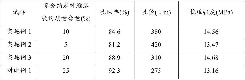
Embodiment 2-3 and comparative example 1

[0051] A bone defect cavity filling implant. Compared with the example, the difference is that in step S1, the mass content of the composite nanofiber solution is shown in Table 1, and the others are roughly the same as in Example 1. In This will not be repeated here.

[0052] Preparation conditions and performance parameters of table 1 embodiment 1-3 and comparative example 1

[0053]

[0054] It can be seen that when the mass content of the nanocomposite fiber solution is too high, the compressive strength decreases. This may be due to the increase of water content in the reaction system, which makes the microwave absorb heat too fast, and the reaction is uneven, resulting in uneven crosslinking structure.


PUM

PropertyMeasurementUnit
Diameteraaaaaaaaaa
Lengthaaaaaaaaaa
Aperture sizeaaaaaaaaaa

Abstract

The invention provides a bone defect cavity filling implant. The bone defect cavity filling implant is of a porous cross-linked network structure formed by chitosan and polylactic acid composite nanofibers and nano-hydroxyapatite growing on the surfaces of the composite nanofibers in series through a sol-gel method. Calcium nitrate and ammonium phosphate are adsorbed to the surfaces of the chitosan and polylactic acid composite nanofibers respectively, and then a sol-gel method is adopted for preparing a porous cross-linked network structure with nano-hydroxyapatite distributed inside in a penetrating mode. In the process, the calcium nitrate and the ammonium phosphate react to obtain the nano-hydroxyapatite, and besides, the composite nanofibers are cross-linked to obtain a gel product. The structure has good biocompatibility, degradability and high mechanical strength, and bone defect healing can be accelerated.

Description

technical field [0001] The invention relates to the technical field of biopolymer material tissue engineering scaffold preparation, in particular to a bone defect cavity filling implant. Background technique [0002] Bone belongs to the supporting structure of the human body and is a part of the human movement system. Its function is to exercise, support and protect the body, and it is of great significance to maintaining human health. Trauma, tumor, surgery, osteoporosis and other factors can cause bone defects. Most of the defective bone tissue can rely on the body's own repair ability to heal the defect site, and return to normal without scar formation. However, when the bone defect area is too large, exceeding the size of the critical bone defect, the bone defect cannot heal itself in the body, and it is necessary to find a suitable bone substitute material to repair the bone defect in order to reconstruct the bone defect, so the repair of large area bone defect is the m...

Claims


Application Information

Patent Timeline
IPC IPC(8): A61L27/12A61L27/18A61L27/20A61L27/50A61L27/56A61L27/58A61F2/28A61F2/30
CPCA61F2/28A61F2/30771A61F2002/2835A61F2002/30784A61L27/12A61L27/18A61L27/20A61L27/50A61L27/56A61L27/58A61L2430/02C08L5/04C08L67/04
Inventor 仇申强
Owner SHANDONG PROVINCIAL HOSPITAL AFFILIATED TO SHANDONG FIRST MEDICAL UNIVERSITY
Why Eureka
Try Eureka