
An RNA fluorescent probe that rapidly differentiates cancer from normal tissue using nucleolus morphological changes

A fluorescent probe and morphological change technology, applied in the field of fluorescent probes, can solve the problems of inability to clearly give the overall outline of the nucleolus, unpublished molecular structure, poor staining selectivity, etc., and achieve the effect of rich details

Active Publication Date: 2021-08-27
SHANDONG UNIV
View PDF4 Cites 0 Cited by
  • Summary
  • Abstract
  • Description
  • Claims
  • Application Information

AI Technical Summary

Problems solved by technology

However, due to the poor selectivity of staining, the overall outline of the nucleolus cannot be clearly given, so the image interpretation related to the nucleolus depends entirely on the doctor's experience
Unfortunately, although there are many RNA fluorescent probes that can image RNA and nucleoli in isolated living cells cultured in vitro, their application in tissues is not ideal
SYTO RNA-Select, as the only commercially available dye for RNA imaging in live cells, not only has its molecular structure unpublished, but its utility in tissues has not been widely demonstrated

Method used


Image

  • An RNA fluorescent probe that rapidly differentiates cancer from normal tissue using nucleolus morphological changes
  • An RNA fluorescent probe that rapidly differentiates cancer from normal tissue using nucleolus morphological changes
  • An RNA fluorescent probe that rapidly differentiates cancer from normal tissue using nucleolus morphological changes

Examples


Embodiment 1

[0040] Example 1: Synthesis of probe CAPY-AP.

[0041]

[0042] The specific synthesis is as follows:

[0043] Synthesis of compound 1: 4-methylpyridine (0.93g, 10mmol) and 3-bromopropylamine hydrobromide (2.41g, 11mmol) were dissolved in 20mL of ethanol, and heated under reflux at 85°C for 12 hours. After the reaction, a white solid appeared. After filtering and washing with petroleum ether, pure white compound 1 was obtained. Yield 70%. 1 H NMR (300MHz, DMSO-d 6 ), δ(ppm): 8.938(d, J=6Hz, 2H), 8.047(d, J=6.3Hz, 2H), 7.797(s, 3H), 4.628-4.582(t, J=6.9Hz, 2H) ,2.860-2.794(m,2H),2.622(s,3H),2.220-2.124(m,2H).

[0044] Synthesis of compound 2: under ice bath condition, POCl 3 (9.2mL, 100mmol) was slowly added to dry DMF (7.7mL, 100mmol), and it became viscous after stirring for 30 minutes. Then dissolve in CH 3 Cl N-ethylcarbazole (2.94g, 10mmol) was added into the system, stirring was continued for 1 hour, and the mixture was heated at 80°C for 12 hours. After cooli...

Embodiment 2

[0046] Example 2: SiHa cell culture.

[0047] Adherently culture SiHa cells in culture medium containing 10% fetal bovine serum at 37°C, 5% CO 2 cultured in a saturated humidity incubator, and the medium was changed every 2-3 days for passage. When the cells grow to the logarithmic phase, culture the slices: ① Soak the coverslips in absolute ethanol for 30 minutes, dry them with an alcohol lamp and place them in a disposable 35mm culture dish for later use; Wash the cells three times with PBS, digest with 1mL 0.25% trypsin for 3-5 minutes, pour out the trypsin carefully, add fresh culture medium and pipette evenly, and count the cells. The concentration is 1×10 per ml 5 , and then inoculated into the above-mentioned petri dish containing the coverslip in 5% CO 2 Cultured in an incubator to allow the cells to grow on the sheet. After the SiHa cells grow on the slide and cover the glass, it is used for the experiment.

Embodiment 3

[0048] Example 3: Staining observation of active SiHa cells by probe CAPY-AP.

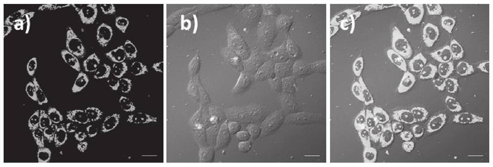
[0049]First prepare 5mM probe CAPY-AP in DMSO solution as mother solution. After the SiHa cells were covered with coverslips, remove the medium in the culture dish, wash the coverslips containing SiHa cells with clean PBS for 3 times, stain the cells with 5 μM CAPY-AP, and store in CO 2 Incubate in the incubator for 30min. Take out the stained slides, wash away the excess probes, cover the cell growth side down on the glass slide, and observe the coloring position, fluorescence distribution and brightness of the cells under a laser scanning confocal fluorescence microscope (using 488nm laser to irradiate the cells) changes etc.

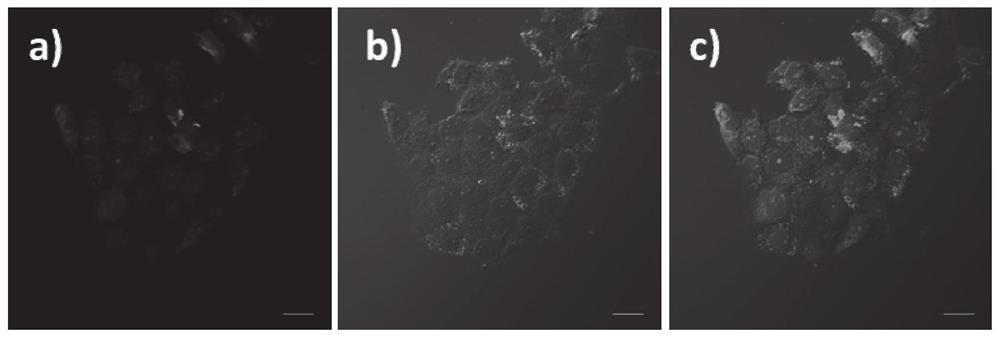
[0050] see results figure 1 . Among them, figure a is the photo of the probe CAPY-AP under excitation of 488nm, collecting the green channel of 500-600nm, figure b is the differential interference micrograph of bright-field laser scanning, and figure c is the merged fi...


PUM


Abstract

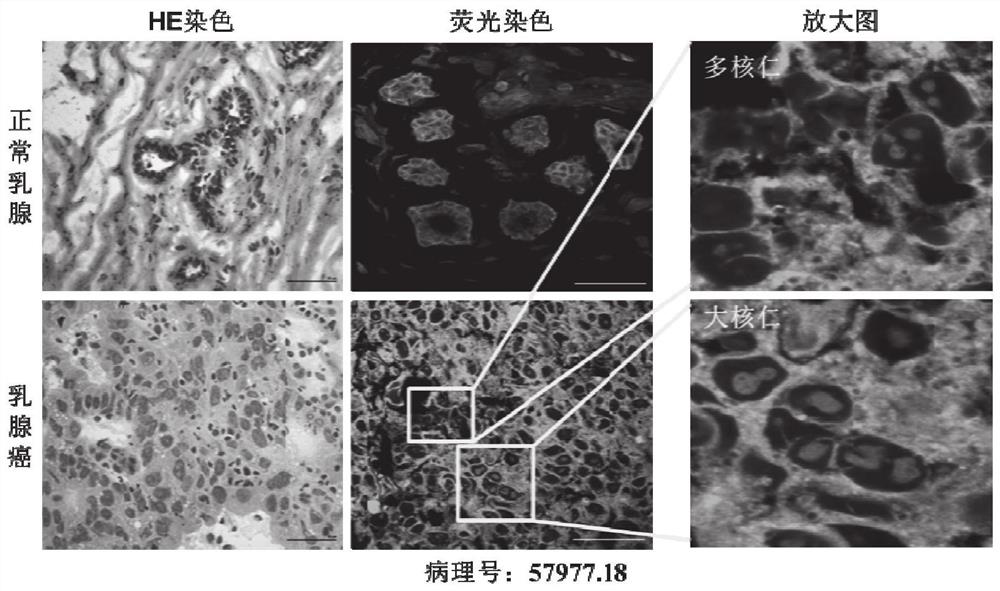
The invention discloses an RNA fluorescent probe for rapidly distinguishing cancer and normal tissue by using nucleolus morphological changes. The probe is (E)-1-(3-aminopropyl)-4-(2-(9-B Base-carbazole-3-alkenyl) vinyl) pyridine dibromide, referred to as CAPY-AP; the RNA fluorescent probe can simultaneously display the distribution of RNA and nucleoli in living cells and normal tissues or cancerous tissues, and the The judgment index for quickly distinguishing cancer from normal tissue by using nucleolus morphology changes is that there is only a single inconspicuous nucleolus in normal tissue, while there are multiple nucleoli and large nucleoli in cancer tissue; compared with other existing RNA probes, The probe of the present invention has high RNA affinity and ultra-high permeability, and can easily image RNA and nucleolus in tissue sections. In addition, the probe of the present invention has the characteristics of good membrane permeability, strong color development, and strong photostability, and is expected to be further applied in the preparation of reagents for rapid pathological diagnosis during tumor surgery.

Description

technical field [0001] The present invention relates to a fluorescent probe capable of simultaneously displaying RNA in living cells and normal tissues or cancerous tissues and its application, in particular to an RNA fluorescent probe capable of rapidly distinguishing cancer from normal tissues by using nucleolus morphological changes and its preparation The application of rapid pathological diagnosis reagent in tumor operation. Background technique [0002] Diagnostic reagents, methods and technologies for major diseases have always been the main field of international high-tech competition. my country is a country with a large population, and the incidence rate of tumors among residents remains high all year round. On average, more than ten thousand people are diagnosed with tumor diseases every day, and many of them are diagnosed with malignant tumors (cancers). According to the latest statistics in January 2019, in 2015, an average of more than 10,000 people were diagn...

Claims


Application Information

Patent Timeline
Patent Type & Authority Patents(China)
IPC IPC(8): C07D401/06C09K11/06G01N21/64
CPCC07D401/06C09K11/06G01N21/6486C09K2211/1029G01N33/582G01N33/57496C12Q2600/156C12Q1/6886C09K2211/1018G01N1/30G01N21/6428G01N2001/302G01N2021/6439
Inventor 于晓强高鹏何秀全孟芳芳贺峻祎刘志强
Owner SHANDONG UNIV
Learn More