Fluorescent biosensor for detecting mercury ions

A biosensor, mercury ion technology, applied in the field of biosensors, can solve the problems of convenience, speed, sensitivity, complex sample pretreatment, and high detection cost that are difficult to adapt to mercury ion detection, and achieve fast detection speed, low detection limit, Fast response time

Active Publication Date: 2021-02-09
UNIV OF JINAN
View PDF5 Cites 5 Cited by
  • Summary
  • Abstract
  • Description
  • Claims
  • Application Information

AI Technical Summary

Problems solved by technology

Currently reported detection methods for mercury ions include atomic emission spectrometry, atomic absorption spectrometry, spectrophotometry, hydride generation-atomic fluorescence spectrometry, etc. Expensive and other issues, it has been difficult to meet the requirements of convenience, speed and sensitivity of mercury ion detection

Method used


Image

  • Fluorescent biosensor for detecting mercury ions
  • Fluorescent biosensor for detecting mercury ions
  • Fluorescent biosensor for detecting mercury ions

Examples


Embodiment 1

[0038] Example 1 Preparation of Composite Probe Q

[0039] (1) Synthesizing S1, S2 and L according to the sequences shown in SEQ ID No: 1-3;

[0040] (2) Dilute sterilized water (36 µL), 10×PBS buffer (pH7.4) (6 µL), S1 (6 µL, 100 µM), S2 (6 µL, 100 µM) and L (6 µL, 100 μM) was added to the pre-prepared sterilized EP tube, shaken for 20-30s, then incubated at 95°C for 5min, cooled slowly to room temperature and hybridized, and stored at -20°C for subsequent experiments.

Embodiment 2

[0041] Example 2 Preparation of cyclic template CP

[0042] (1) Synthesize 5' phosphorylated padlock probe TP and ligation probe LP according to the sequences shown in SEQ ID No: 5 and 6;

[0043] (2) Mix TP and LP with equal volume and molar concentration (6 µL, 100 µM), add 10×T4 DNA ligase buffer (6 µL) and incubate at 95°C for 5 min, cool to room temperature, then add T4 DNA Ligase (3 µL, 400 U / µL), incubated overnight at 16°C, then heated at 65°C for 10 min to inactivate the enzyme; add Exo I enzyme (2 µL, 20 U / µL) and Exo III enzyme (1 µL, 100 U / µL) was digested at 37°C for 2 h, then heated at 85°C for 20 min to inactivate the enzyme to obtain the circular template CP with the sequence shown in SEQ ID No: 4, and finally stored at 0-4°C for subsequent experiments .

Embodiment 3

[0044] Example 3 Trigger chain T concentration screening

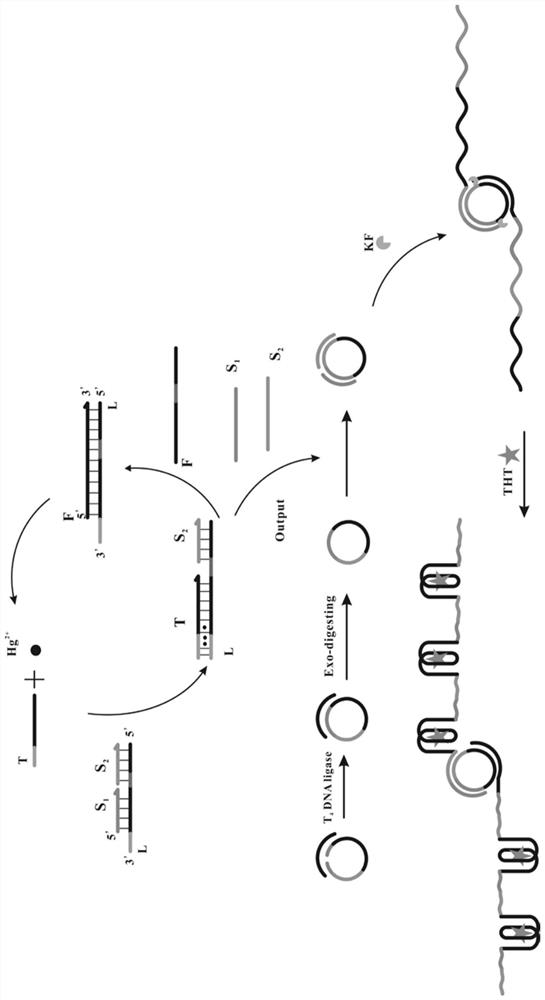
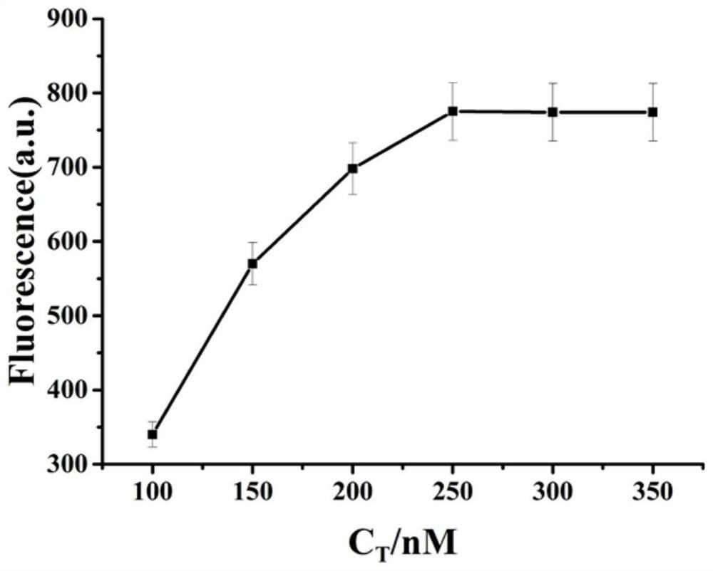
[0045](1) Dilute sterilized water (6 µL), 10× buffer 2 (3 µL), Hg 2+ (3 µL, 10 µM), trigger chain T (3 µL, make the final concentration 100nM, 150nM, 200nM, 250nM, 300nM, 350nM), composite probe Q (3 µL, the final concentration is 2 µM), fuel chain Add F (3 µL), circular template CP (3 µL, 1 µM), KF (3 µL, final concentration 0.5 U / µL) and ThT (3 µL, final concentration 10 µM) into the centrifuge tube, shake 20-30s, in a water bath at 37°C for 2h, the product was obtained;

[0046] (2) Dilute the above product to 100 µL, and measure the fluorescence intensity at 485 nm with 425 nm as the excitation wavelength;

[0047] see results figure 2 , it can be seen from the figure that with the increase of the concentration of the trigger chain T, the fluorescence intensity obtained in the experiment is continuously enhanced. is 250nM.

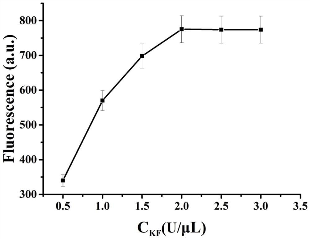
[0048] Example 3 KF concentration screening

[0049] (1) Dilute sterilized water (6 ...


PUM


Abstract

The invention provides a fluorescent biosensor for detecting Hg<2+>. The fluorescent biosensor comprises a composite probe Q, an annular template CP, a trigger chain T, a fuel chain F, a Klenow fragment and thioflavin T; and the trigger chain T, the composite probe Q, the fuel chain F, the annular template CP and the Klenow fragment are uniformly mixed with a mercury ion solution with a series ofconcentrations and a to-be-detected solution respectively, incubating is performed, enzyme is inactivated, the sulfur element T is added, incubating is performed, and the fluorescence intensity is detected. Target cyclic amplification and signal amplification are realized on the basis of the specific binding capacity between basic groups T and Hg<2+> and entropy-driven strand displacement amplification and rolling circle amplification reactions, and the biosensor for sensitive detection of a target object is realized. The sensor has the advantages of high detection speed, low detection limit,high sensitivity and the like, can make up the defects and deficiencies of the existing detection method of Hg<2+>, and realizes rapid and accurate quantitative detection of Hg<2+>.

Description

technical field [0001] The invention belongs to the technical field of biosensors, and relates to a fluorescent biosensor for detecting mercury ions. Background technique [0002] Mercury is one of the potentially dangerous and toxic elements. Because of its bioaccumulation, even a very small amount of mercury is highly toxic to human health and the environment. In recent years, with the development of industry, mercury ion pollution has become increasingly serious, causing great harm to human beings and the environment. Metallic mercury poisoning is often caused in the form of mercury vapor. Because mercury vapor is highly diffusible and fat-soluble, it enters the alveoli through the respiratory tract and is transported to the whole body through blood circulation. After the metallic mercury in the blood enters the brain tissue, it is oxidized into mercury ions and gradually accumulates in the brain tissue. When it reaches a certain amount, it will cause damage to the brai...

Claims


Application Information

Patent Timeline
Patent Type & Authority Applications(China)
IPC IPC(8): G01N21/64C12Q1/682C12Q1/6825
CPCG01N21/64G01N21/6428G01N21/643C12Q1/682C12Q1/6825G01N2021/6417G01N2021/6439C12Q2521/501C12Q2531/119C12Q2531/125C12Q2563/107C12Q2565/607
Inventor 张曼茹刘素黄加栋王玉江龙孙文玉王业茹张清心徐婉晴朱志学李静静
Owner UNIV OF JINAN
Why Eureka
Try Eureka