
Nanoparticle composite showing improved endocytosis efficiency through surface modification using lipid and manufacturing method therefor

A nanoparticle and manufacturing method technology, applied in the direction of liposome delivery, other methods of inserting foreign genetic materials, medical preparations of non-active ingredients, etc., can solve the problems of decreased stability and insufficient achievement, and achieve large-scale The effect of mass production

Active Publication Date: 2021-02-26
IND UNIV COOP FOUND SOGANG UNIV
View PDF9 Cites 1 Cited by
  • Summary
  • Abstract
  • Description
  • Claims
  • Application Information

AI Technical Summary

Problems solved by technology

[0005] However, the existing techniques for improving the efficiency of intracellular uptake cannot achieve sufficient effects, and there is a problem of decreased stability due to the easy decomposition of the bound substances

Method used


Image

  • Nanoparticle composite showing improved endocytosis efficiency through surface modification using lipid and manufacturing method therefor
  • Nanoparticle composite showing improved endocytosis efficiency through surface modification using lipid and manufacturing method therefor
  • Nanoparticle composite showing improved endocytosis efficiency through surface modification using lipid and manufacturing method therefor

Examples


Embodiment 1

[0056] Production of Nanoparticle Composite

[0057] 1. Formation of Nanoparticles (NPs)

[0058] After dissolving albumin (Human serum albumin) in distilled water to a concentration of 20mg / mL, use 0.2M NaOH to adjust the pH to 8 to prepare an albumin solution, and then add 100% ethanol to the above albumin solution at a rate of 1mL / min Titrate. Next, 10 μL of 4% glutaraldehyde (glutaraldehyde) was added and ethanol was evaporated overnight under light-shielding conditions, and after centrifugation was performed at 13200 rpm for 10 minutes, unparticled albumin was removed with a pipette, Next, use a pipette to obtain the supernatant (nanoparticles (NPs)) except micropellets (micropellets) after redispersing with phosphate buffered saline (PBS) and performing centrifugation under the conditions of 3000rpm and 5min (in addition, When performing the fluorescence test, react the fluorescent dye (dye) and nanoparticles that meet the requirements overnight at room temperature, t...

Embodiment 2

[0066] Characteristic Confirmation of Nanoparticle Complex

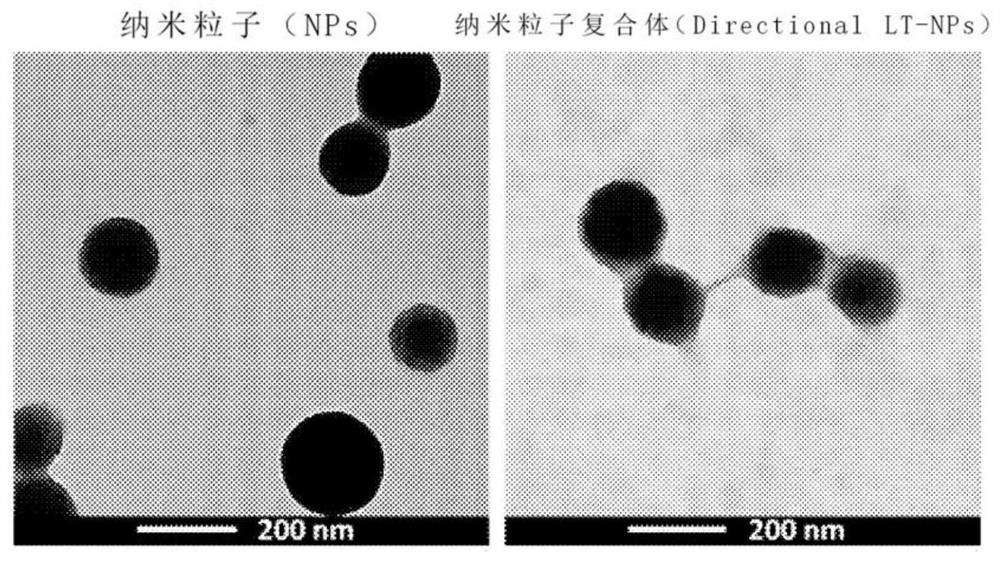
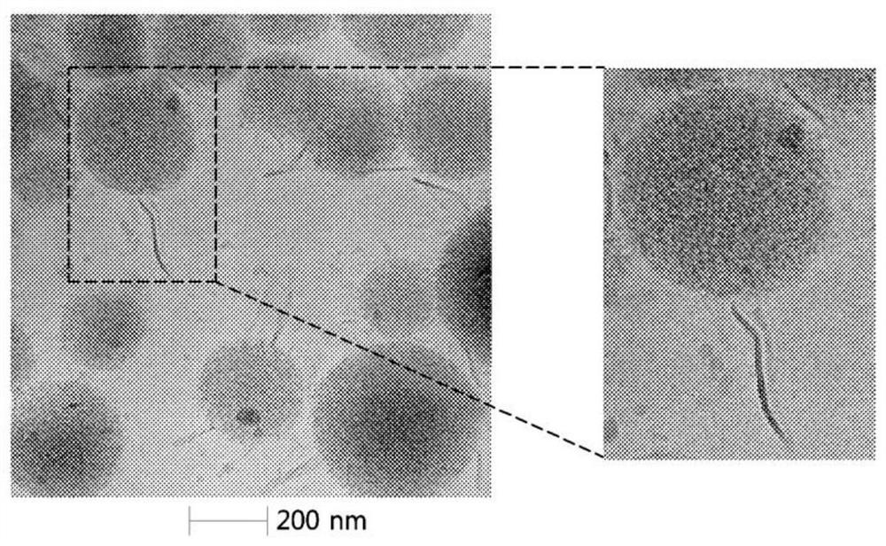
[0067] 1. Utilize transmission electron microscope (TEM) to measure the nanoparticles (NPs) formed in 1 of embodiment 1 and the nanoparticle complex (Directional LT-NPs) formed in (2) in 4 of embodiment 1 , the result is as figure 2 Shown, and utilize frozen transmission electron microscope (Cyro-TEM) to measure above-mentioned nanoparticle complex and lipid structure body, its result is as follows respectively image 3 as well as Figure 4 shown.

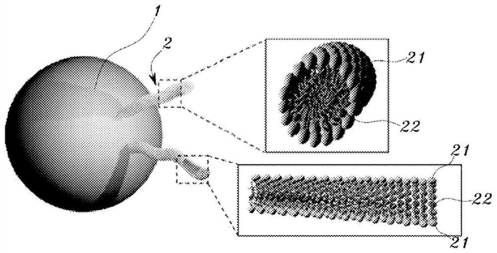
[0068] 2. pass figure 2 It was confirmed that the surface of the nanoparticle was smooth, but the nanoparticle complex showed an uneven surface due to the lipid structure attached to the surface of the nanoparticle. Additionally, by image 3 It can be confirmed that a tubular lipid structure is attached to the surface of the nanoparticle, by Figure 4 It was clearly confirmed that the above-mentioned lipid structure had a tubular form. Additionally, by image 3...

Embodiment 3

[0069] Evaluation of Cellular Uptake Efficiency of Nanoparticle Complex

[0070] 1. In order to evaluate the cellular uptake efficiency of the nanoparticle (NPs) formed in 1 of Example 1 and the nanoparticle complex (Directional LT-NPs) formed in (2) of Example 1 Flow cytometry (Flowcytometry) for analysis, the results are as follows Figure 5 shown, and imaged using a confocal microscope, the results are shown in Image 6 shown. The analysis by flow cytometry is performed by using Alexa 488 fluorescent dye (dye) labeled nanoparticles and nanoparticle complexes on A549 cells (1 × 10 4 ), while the analysis using confocal microscopy was performed by using Cy5.5 fluorescent dye (dye)-labeled nanoparticles and nanoparticle complexes to stain nuclei with DAPI and use phalloidin (phalloidin) A549 cells stained for cytoskeleton (1×10 5 ) is executed in the manner of processing.

[0071] 2. In addition, in order to compound the nanoparticles (NPs) formed in 1 of Example 1 and t...


PUM

PropertyMeasurementUnit
diameteraaaaaaaaaa
lengthaaaaaaaaaa
widthaaaaaaaaaa

Abstract

The present invention relates to a nanoparticle composite, which is endocytosed into cells and used to treat a disease, and to a manufacturing method therefor. More specifically, the present inventionrelates to a nanoparticle composite and a manufacturing method therefor, wherein the nanoparticle composite shows improved endocytosis efficiency by modifying the nanoparticle surface with a lipid-based material having high stability and excellent biocompatibility; the nanoparticle composite can attain direct passage through the cellular membrane as well as endocytosis, and can be effectively endocytosed into spheroid-shaped tumor cells, by having a tube-shaped lipid structure combined with a portion of the nanoparticle surface; and the nanoparticle composite can be easily mass-produced by using a top-down manner, in which, the lipid structure is not attached directly to the nanoparticle, but the binding of the nanoparticle and the lipid-based lipidome (bubble, liposome, etc.) is induced,and then the physical force is applied thereto to disrupt the lipidome, thereby forming the lipid structure on the nanoparticle surface.

Description

technical field [0001] The present invention relates to a nanoparticle complex used for treatment of diseases by being taken up into cells and a method for producing the same, and particularly relates to a lipid-based substance that can be used to treat diseases with high stability and excellent biocompatibility Modifying the surface of nanoparticles to improve the efficiency of intracellular uptake can achieve endocytosis and even directly penetrate the cell membrane by combining a tubular lipid structure on the surface of nanoparticles, and can also be effectively absorbed by tumor cells in the form of spheroids. uptake, and can be broken up by applying mechanical force after binding lipid-based liposomes (such as microvesicles, membrane vesicles, etc.) to nanoparticles and thereby forming lipid structures on the surface of nanoparticles A nanoparticle complex and a manufacturing method thereof that can be easily mass-produced in a top-down manner. Background technique ...

Claims


Application Information

Patent Timeline
Patent Type & Authority Applications(China)
IPC IPC(8): A61K9/50A61K9/51A61K9/127A61K47/69A61K45/06
CPCA61K9/127A61K31/7105A61K31/712A61K31/704A61K9/5015A61K9/1271A61K9/1277A61K9/5089A61K47/6919A61K47/6929A61K47/6911A61K47/6925C12N15/88A61K9/5192A61K9/5123A61K45/06
Inventor 金铉喆金度延
Owner IND UNIV COOP FOUND SOGANG UNIV
Learn More