A structural color microfiber with heterogeneous structure and its preparation method and cardiomyocyte detection method

A technology of heterogeneous structures and cardiomyocytes, applied in the fields of fiber chemical characteristics, chemical instruments and methods, and measuring devices, can solve the problems of lack of effective in vitro models for new cardiac drugs and dependence on cardiomyocyte sensing, and achieve more controllable optical properties , high sensing stability and sensitivity, and the effect of ensuring stability

Active Publication Date: 2022-04-26
NANJING DRUM TOWER HOSPITAL
View PDF4 Cites 0 Cited by
  • Summary
  • Abstract
  • Description
  • Claims
  • Application Information

AI Technical Summary

Problems solved by technology

[0005] In order to solve the shortcomings of myocardial cell sensing relying on large and complex instruments and the lack of effective in vitro models for the development of new cardiac drugs, the present invention provides a microfluidic chip-based structured color microfiber with axial heterogeneous structure and its preparation method and Cardiomyocyte detection method, said cardiomyocyte detection method is characterized in that the periodic contraction of cardiomyocytes pulls the microfiber structural color area to produce deformation and color changes, and realizes the visual detection and sensing of cardiomyocytes

Method used


Image

  • A structural color microfiber with heterogeneous structure and its preparation method and cardiomyocyte detection method
  • A structural color microfiber with heterogeneous structure and its preparation method and cardiomyocyte detection method
  • A structural color microfiber with heterogeneous structure and its preparation method and cardiomyocyte detection method

Examples


Embodiment 1

[0031] This example provides a preparation method of a two-component heterogeneous structural color microfiber and a method for detecting cardiomyocytes based on single-end stretching.

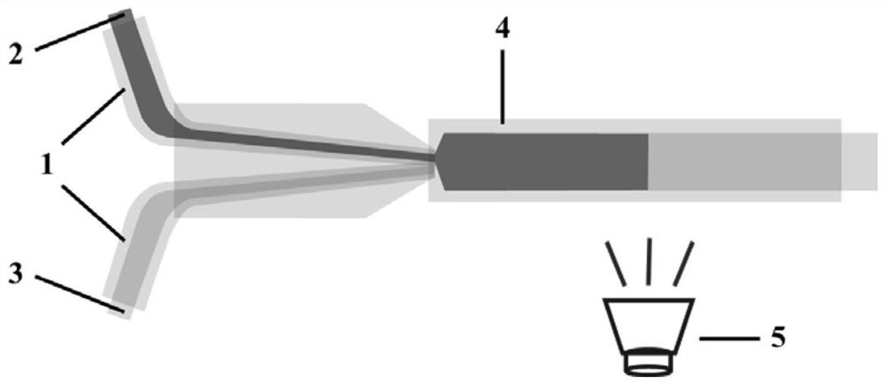
[0032] The preparation method process is as figure 1 Shown: Use a hydrophobic capillary with an inner diameter of 200 μm as the collection tube. Heat and elongate the capillary tube with an inner diameter of 300 μm in the outer flame of the spray gun to form a tapered front end. Cut off two sections of tapered front end capillary tubes of appropriate length as the liquid injection port. The tapered front end of the liquid injection port is connected to the collection pipe. Assembled into a microfluidic chip.

[0033] Configure a 15% (w / v) aqueous solution of methacrylated gelatin (GelMA), and disperse sulfonate-modified silica nanoparticles (particle diameter about 145 nm) in a concentration of 12% (w / v) in 15% (w / v) Polyacrylamide (AAm):N,N'-methylenebisacrylamide (Bis, crosslinking agent) ...

Embodiment 2

[0039] This example provides a preparation method of a two-component heterogeneous structural color microfiber and a method for detecting cardiomyocytes based on single-end stretching.

[0040] Preparation method: Use a hydrophobic capillary with an inner diameter of 300 μm as a collection tube. Heat and elongate the capillary tube with an inner diameter of 500 μm in the outer flame of the spray gun to form a tapered front end, cut off 4 sections of tapered front end capillary tubes with a suitable length as the liquid injection port, and the tapered front end of the liquid injection port is connected to the collection pipe. Assembled into a microfluidic chip.

[0041] Configure 20% (w / v) GelMA aqueous solution; polyethylene glycol diacrylate (PEGDA) and AAm (containing 1 / 30 cross-linking agent Bis) are prepared in a ratio of 2:15 to form a composite hydrogel prepolymer Body solution, sulfonate-modified silica nanoparticles (particle size about 145nm) were dispersed in 15% (w...


PUM

PropertyMeasurementUnit
diameteraaaaaaaaaa
lengthaaaaaaaaaa

Abstract

The invention discloses a structurally colored microfiber with a heterogeneous structure, a preparation method thereof, and a detection method for cardiomyocytes. Polymerization solution is used as fiber component, and prepolymerization solution includes biocompatible hydrogel solution and at least one concentration of silica non-close-packed colloidal crystal pregel, and a photoinitiator is added to each prepolymerization solution; The prepolymer-like solution is programmed and alternately injected into the collection tube through the microfluidic chip, and the collection tube is continuously irradiated with ultraviolet light to obtain structurally colored microfibers with a heterogeneous structure. The invention has the advantages of controllable preparation, low cost, and can realize visualized quantitative sensing of cell physiology and the like.

Description

technical field [0001] The invention belongs to the field of biomedical materials, and in particular relates to a structurally colored microfiber with a heterogeneous structure, a preparation method thereof, and a cardiomyocyte detection method. Background technique [0002] Cell-generated mechanical forces play an important role in regulating biological functions from cell to tissue level. The abnormality of cell mechanical force is usually related to the dysfunction of organ tissue and abnormal anatomical state, so cell mechanical force has gradually been regarded as an element of disease assessment. To effectively measure cellular mechanical forces, researchers have proposed and developed various mechanical force sensing techniques, such as atomic force microscopy, traction force microscopy, elastomeric micropillar array analysis, etc. Although these micromechanical sensing technologies have great advantages in the observation of biomechanical processes and subcellular s...

Claims


Application Information

Patent Timeline
Patent Type & Authority Patents(China)
IPC IPC(8): G01N21/78G01B11/16D01F6/44D01F1/10B01L3/00B01J19/12
CPCG01N21/78G01B11/16D01F6/44D01F1/10B01L3/502707B01J19/123
Inventor 赵远锦陈卓玥王月桐池俊杰张大淦商珞然
Owner NANJING DRUM TOWER HOSPITAL
Why Eureka
Try Eureka