Application of HSP70 as molecular marker to detection of thalassemia and preparation of diagnostic kit
A thalassemia and molecular marker technology, applied in the application field of preparing diagnostic kits, can solve the problems of cumbersome operation steps, inability to make accurate judgments, long diagnosis time, etc., and achieve good specificity, diversified detection techniques, and convenient detection. Effect
- Summary
- Abstract
- Description
- Claims
- Application Information
AI Technical Summary
Problems solved by technology
Method used
Image
Examples
Embodiment 1
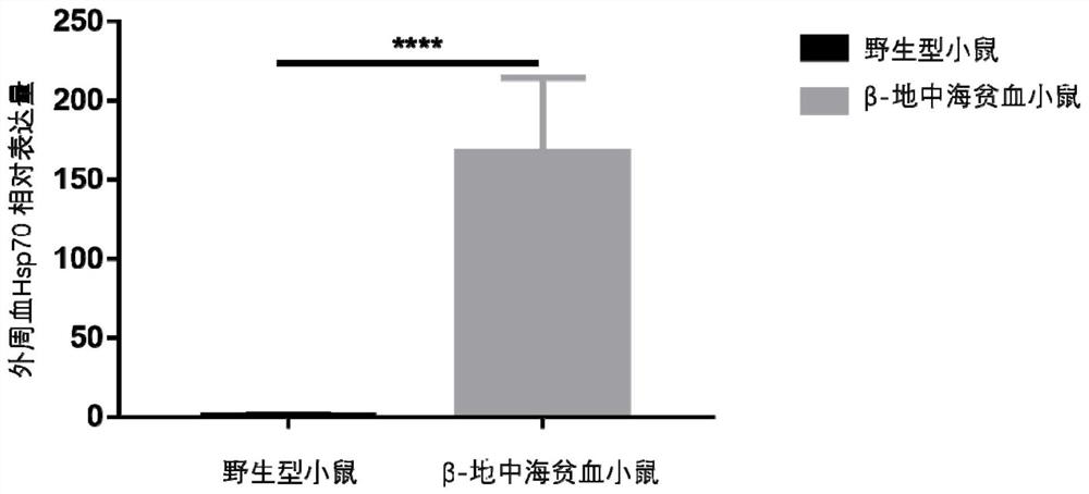
[0020] Example 1: In this example, qRT-PCR technology was used to detect the mRNA level of HSP70 in peripheral blood of mice.
[0021] Experimental steps:
[0022] 1) Collect peripheral blood of mice with anticoagulant EP tube;
[0023] 2) Add 1ml RNA extraction reagent (Trizol) solution to lyse red blood cells and extract total RNA; μ
[0024] 3) Detect the concentration of total RNA and reverse transcribe it into cDNA;
[0025] 4) Configure 10 μL of qPCR reaction system, as follows:
[0026] cDNA (30ng, 2μL); SYBR Green fluorescent dye (5μL); double distilled water (1μL); HSP70 or internal reference gene GAPDH (glyceraldehyde-3-phosphate dehydrogenase) primer (10mM, 2μL). The HSP70 forward primer sequence is 5'-CCATCGAGGAGGTGGATTAGA-3', as shown in SEQ ID NO.1; the HSP70 reverse primer sequence is 5'-AGTGCTGCTCCCAACATTAC-3', as shown in SEQ ID NO.2; GAPDH forward The primer sequence is 5'-ATCATCCCTGCATCCACT-3', as shown in SEQ ID NO.3; the GAPDH reverse primer sequence i...
Embodiment 2
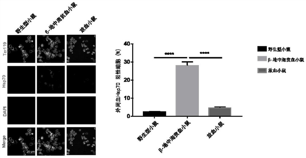
[0029] Example 2: In this example, immunofluorescence was used to detect the expression level of HSP70 in peripheral blood of mice.
[0030] Experimental steps:
[0031] 1) Collect peripheral blood of mice with anticoagulant EP tube;
[0032] 2) Take 10 μl of anticoagulated blood and slowly add 1ml of ice-methanol for internal fixation for 20 minutes;
[0033] 3) After the fixation, evenly smear the blood cells on the glass slide;
[0034] 4) Permeabilize the membrane with 0.1% Triton X-100 by volume for 10 minutes, and wash 3 times with pH7.2-7.4 phosphate buffer saline (PBS);
[0035] 5) blocking with bovine serum albumin at a mass concentration of 1% for 30 minutes;
[0036] 6) HSP70 primary antibody (Abcam, USA) was incubated overnight at 4°C;
[0037] 7) Recover the antibody, wash it with PBS for 3 times, add the antibody of erythroid cell surface marker glycoprotein Ter119 (Biolegend, USA), and the goat anti-rabbit secondary antibody labeled with fluorescein isothioc...
Embodiment 3
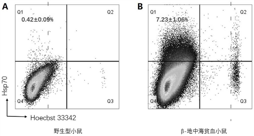
[0041] Example 3: In this example, flow cytometry was used to detect the expression level of HSP70 in peripheral blood of mice.
[0042] Experimental steps:
[0043] 1) Collect peripheral blood of mice with anticoagulant EP tube;
[0044] 2) Take 10 μl of anticoagulated blood and slowly add 1ml of ice-methanol for internal fixation for 20 minutes;
[0045] 3) Wash the cells once, and permeabilize the membrane with 0.1% Triton X-100 for 10 minutes;
[0046] 4) After waiting for natural sedimentation, discard the supernatant, then add HSP70, Ter119, Hoechst 33342 flow-type antibodies and incubate in the dark for 20 minutes;
[0047] 5) After the incubation, the cells were washed and detected by a flow cytometer.
[0048] Experimental results such as image 3 Shown: the proportion of HSP70 positive cells detected by flow cytometry in peripheral blood cells of wild-type mice (Ter119 positive and Hoechst3342 negative) was 0.42 ± 0.09% ( image 3 Middle A), while thalassemia mi...
PUM
 Abstract
Description
Claims
Application Information
 - R&D
- Intellectual Property
- Life Sciences
- Materials
- Tech Scout
- Unparalleled Data Quality
- Higher Quality Content
- 60% Fewer Hallucinations
Browse by: Latest US Patents, China's latest patents, Technical Efficacy Thesaurus, Application Domain, Technology Topic, Popular Technical Reports.
© 2025 PatSnap. All rights reserved.Legal|Privacy policy|Modern Slavery Act Transparency Statement|Sitemap|About US| Contact US: help@patsnap.com


