Polypeptide imaging probe as well as preparation method and application thereof

A technology of imaging probes and peptide segments, which is applied in the fields of peptide preparation, fusion peptides, chemical instruments and methods, etc., can solve the problems of inefficient access, limited application scope of imaging probes, and large molecular weight of probes, and achieves structural Simple, good aggregation retention, and the effect of enhancing the imaging signal-to-noise ratio

Active Publication Date: 2021-05-14
THE NAT CENT FOR NANOSCI & TECH NCNST OF CHINA
View PDF4 Cites 0 Cited by
  • Summary
  • Abstract
  • Description
  • Claims
  • Application Information

AI Technical Summary

Problems solved by technology

However, the imaging probe has an enzyme-substrate unit, which makes the molecular weight of the probe large and cannot enter cells efficiently, and the substrate needs to react with a high concentration of enzyme around the lesion to trigger the self-assembly of the probe, which limits the applicability of the imaging probe scope

Method used


Image

  • Polypeptide imaging probe as well as preparation method and application thereof
  • Polypeptide imaging probe as well as preparation method and application thereof
  • Polypeptide imaging probe as well as preparation method and application thereof

Examples


Embodiment 1

[0073]Example 1 Design, Synthesis and Function of Polypeptide Imaging Probe αvβ3-CY

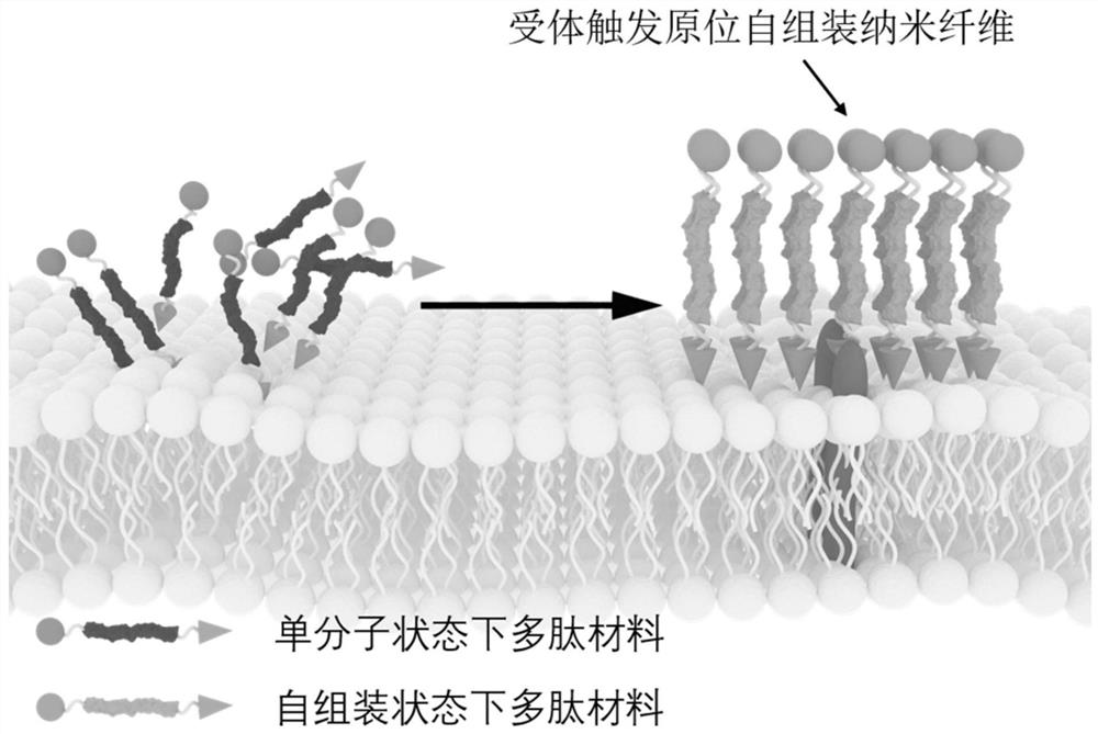
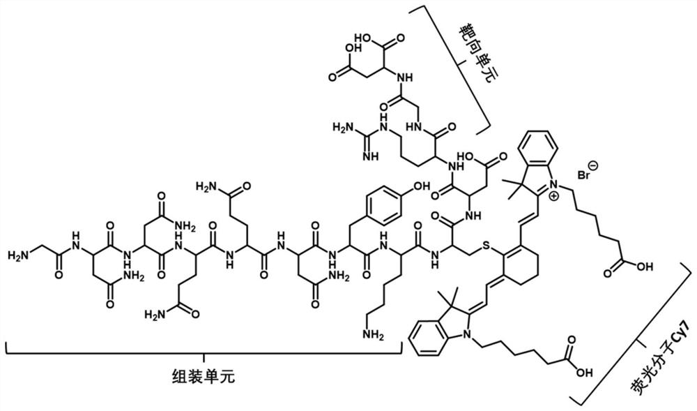
[0074]In this example, the polypeptide imaging probe αvβ3-CY is designed with αvβ3 as a targeted receptor, and the polypeptide imaging probe αvβ3-cyp is made of a peptide, soluble self-contained peptide, and near-infrared fluorescent molecule Cy. Near infrared fluorescent molecule CY is attached to the side chain of polypeptides by cysteine ​​(C), and molecular structural formula isfigure 2 As shown, the amino acid sequence is shown in SEQ ID NO: 14;

[0075]SEQ ID NO: 14: GnnqqNykc (CY7) DRGD.

[0076]The synthesis steps are as follows:

[0077](1) Weighing the resin and invested in the polypeptide solid phase synthesis, adding an appropriate amount of DMF swelling for 4 h; extracting the DMF, perform FMOC to protect by FMOC deprotectant, shake the bed mixed 15 min; extract the FMOC deprotectant, Add DMF, DCM alternately washing the resin 3 times, take a small amount of resin (about 10) from the polypeptide s...

Embodiment 2

[0087]Example 2 Design, Synthesis and Function of Polypeptide Imaging Probes EPCAM-CY

[0088]In this embodiment, the polypeptide imaging probe EPCAM-CY is designed with EPCAM, the polypeptide imaging probe EPCAM-CY consists of an EPCAM receptor recognition peptide, a soluble self-contained peptide and a near-infrared fluorescent molecule Cy. Near infrared fluorescent molecule CY is attached to the side chain of polypeptides by cysteine ​​(C), and molecular structural formula isFigure 5 As shown, amino acid sequences are shown in SEQID NO: 15;

[0089]SEQ ID NO: 15: gnnqqNykc (CY7) Dyevhtyyld.

[0090]Synthesis method is shown in Example 1.

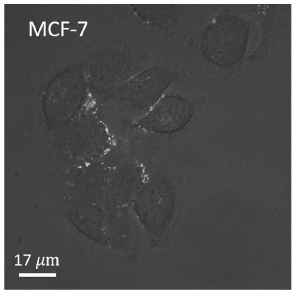
[0091]Around 10 respectively5Epithelial cell adhesion molecule EPCAM high expression of breast cancer cell line MCF-7 and 1051 ml of 20 μm polypeptide imaging probe EPCAM-CY, 5% CO, epstel, umbilical vein endothelial cell line, ephemeric cell adhesion molecule epcam, is added to HUVEC.2The cell incubator was incubated for 1 h, and the cells were observed u...

Embodiment 3

[0096]Example 3 Design, Synthesis and Function of Polypeptide Imaging Probe αvβ3-NBD

[0097]Compared to near-infrared fluorescent molecules, the biographical imaging effect of short-wavelength fluorescent molecules is slightly insufficient, but the cell imaging effect is more stable, and it is not easy to quench. This example employs a fluorescence imaging of the cellular surface with a molecule having a aggregated induced light-emitting effect of an aggregation induced light-emitting effect, having an exemption, signal-to-noise ratio, and can qualitatively characterize the expression of the cell surface receptor.

[0098]The molecular structure formula of the polypeptide imaging probe αvβ3-NBD in this embodiment is likeFigure 8 As shown, amino acid sequences are shown in SEQ ID NO: 16, wherein X is Fmoc aminopteraenic acid;

[0099]SEQ ID NO: 16: NBD-X-gnnqqNyRGD.

[0100]Synthesis Methods Example 1, different in the case where the NBD is counted: FMOC is removed after the last amino acid of ...


PUM


Abstract

The invention provides a polypeptide imaging probe as well as a preparation method and application thereof. The polypeptide imaging probe comprises a receptor recognition peptide fragment, a self-assembly peptide fragment and a signal molecule. According to the polypeptide imaging probe, the receptor recognition peptide fragment is combined with a receptor in a recognition manner, the self-assembly peptide fragment is triggered to generate self-assembly, and the signal molecule sends out a signal. The polypeptide imaging probe is self-assembled to form nanofibers through tumor microenvironment response, compared with a small molecule imaging agent, the polypeptide imaging probe has the advantages of high enrichment and long retention, the polypeptide imaging probe is not easy to metabolize along with time extension, has a long-acting retention effect, presents a high signal-to-noise ratio at a targeted part, and is low in required dosage, so that toxicity to metabolic organs is remarkably reduced, and a new thought and a new method are provided for transformation and development of imaging.

Description

Technical field[0001]The present invention belongs to the field of nano biopharmaceutical technology, which relates to a polypeptide imaging probe and a preparation method thereof, in particular, to an inner self-assembled polypeptide imaging probe in which cell receptor is identified and has a preparation method thereof and / in cell imaging and / Or use in living imaging.Background technique[0002]Malignant tumor (cancer) is a disease that seriously threatens human life. In the clinical treatment of cancer, imaging is used to imaging the tumor lesions using imaging probes, which is advantageous for achieving early diagnosis, lesion positioning, intraoperative navigation and other purposes. However, conventional small molecules such as indoleptic (ICG), methylene blue (MB), etc., has low enriched efficiency, short retention time, weak fluorescent signal-to-noise ratio, and use dose, which may result in false positivity As a result, the metabolic organism is highly poisonous, limitin...

Claims


Application Information

Patent Timeline
IPC IPC(8): C07K19/00C07K1/13C09K11/06C09K11/02G01N21/64A61K49/00
CPCC07K5/0817C07K7/06C07K7/08C09K11/06C09K11/025G01N21/6428G01N21/6458A61K49/0056C07K2319/735C09K2211/1029C09K2211/1048C09K2211/1044G01N2021/6432
Inventor 王浩安红维王曼迪吕甘田
Owner THE NAT CENT FOR NANOSCI & TECH NCNST OF CHINA
Why Eureka
Try Eureka