EGF mesenchymal stem cell exosome as well as preparation method and application thereof

A technology of mesenchymal stem cells and exosomes, applied in the field of EGF mesenchymal stem cell exosomes and its preparation, can solve the problems of insignificant biological activity, inability of EGF to play a specific targeting role, and low content, and achieve Good therapeutic effect, promoting proliferation and migration, accelerating healing effect

Inactive Publication Date: 2021-07-09
广州远想生物科技股份有限公司
View PDF4 Cites 3 Cited by
  • Summary
  • Abstract
  • Description
  • Claims
  • Application Information

AI Technical Summary

Problems solved by technology

And in the damaged site, although the EGF carried by exosomes can also be delivered to the recipient cells to play a role, but because it is expressed in the membrane, EGF cannot play a specific targeting role, and its content is low. The biological activity is not very significant
There is no report on the combination of EGF and exosomes for tissue wound repair

Method used


Image

  • EGF mesenchymal stem cell exosome as well as preparation method and application thereof
  • EGF mesenchymal stem cell exosome as well as preparation method and application thereof
  • EGF mesenchymal stem cell exosome as well as preparation method and application thereof

Examples


Embodiment 1

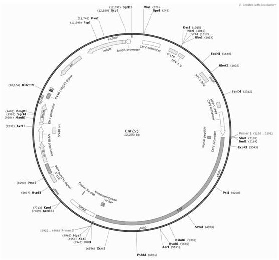
[0039] The construction of embodiment 1 lentiviral vector

[0040] 1. Design the lentiviral vector of the fusion protein EGF, innovatively design the corresponding sequence of the fusion protein according to the signal peptide sequence and transmembrane sequence of the mesenchymal stem cell surface marker CD44, each sequence is as follows: N-terminal signal peptide:

[0041] ATGGACAAGTTTTGGTGGCACGCAGCCTGGGGACTCTGCCTCGTGCCGCTGAGCCTGGCG (SEQ ID NO. 1)

[0042] Target gene EGF:

[0043]ATGCTGCTCACTCTTATCATTCTGTTGCCAGTAGTTTCAAAATTTAGTTTTGTTAGTCTCTCAGCACCGCAGCACTGGAGCTGTCCTGAAGGTACTCTCGCAGGAAATGGGAATTCTACTTGTGTGGGTCCTGCACCCTTCTTAATTTTCTCCCATGGAAATAGTATCTTTAGGATTGACACAGAAGGAACCAATTATGAGCAATTGGTGGTGGATGCTGGTGTCTCAGTGATCATGGATTTTCATTATAATGAGAAAAGAATCTATTGGGTGGATTTAGAAAGACAACTTTTGCAAAGAGTTTTTCTGAATGGGTCAAGGCAAGAGAGAGTATGTAATATAGAGAAAAATGTTTCTGGAATGGCAATAAATTGGATAAATGAAGAAGTTATTTGGTCAAATCAACAGGAAGGAATCATTACAGTAACAGATATGAAAGGAAATAATTCCCACATTCTTTTAAGTGCTTTAAAATATCCTGCAAATGTAGCAGTTGATCCAGT...

Embodiment 2

[0057] Example 2 Characterization of Mesenchymal Stem Cells Infected with Viruses

[0058] 1. For the method of cell infection, take 10ug of the successfully constructed EGF plasmid and add it to the serum-free DMEM medium, then add 30μL of lipo3000 to it, mix well and let stand for 20min, add to the cell number of 4×10 6 HEK 293T cells, mix gently, place at 37°C, 5% CO 2 After 6 hours, the 5% FBS complete medium was replaced with 10 mL of 30% FBS complete medium. After 48 hours, the cell supernatant was collected and centrifuged at 4000 rpm for 15 minutes at room temperature. Take supernatant and add to cell number 4×10 6 Add polybrene with a final concentration of 8ug / ml to the mesenchymal stem cells, mix well, and place at 37°C, 5% CO 2 After 12 hours, replace with 10% FBS complete medium.

[0059] 2. Add puromycin at a final concentration of 2ug / ml to the infected cells after 24 hours to screen for resistant cells.

[0060] 3. Extract the RNA of wild-type mesenchymal c...

Embodiment 3

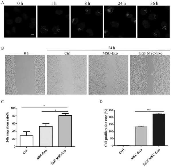
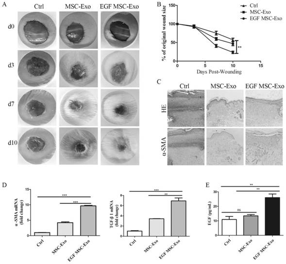
[0070] Example 3 Characterization of exosomes derived from mesenchymal stem cells after infection with lentivirus

[0071] 1. Extract wild-type mesenchymal cells (MSC), mesenchymal stem cells (MSC+EGF) co-incubated with EGF growth factor, and exocytosis of mesenchymal stem cells (EGF MSC) after lentivirus infection with integrated EGF sequence body. After the cells were cultured with 10% FBS complete medium to a density of 70%, they were replaced with 0.5% EVFree FBS medium, and after 48 hours, the cells were harvested on the culture medium. Centrifuge at 4°C, 500×g, 10min, and take the supernatant. Centrifuge at 4°C, 2000×g, 20min, and take the supernatant. Centrifuge at 4°C, 10000×g, 40min, and take the supernatant. Centrifuge at 4°C, 100000×g, 90min, and take the precipitate. After resuspending with PBS, centrifuge at 4°C, 100,000×g, 90min, take the precipitate and get the exosomes of mesenchymal stem cells, resuspend the exosomes with 100μl PBS and store in -80°C refri...


PUM


Abstract

The invention relates to an EGF mesenchymal stem cell exosome, which is secreted by mesenchymal stem cells and can express EGF fusion protein on a mesenchymal stem cell membrane. The EGF fusion protein sequentially comprises an N-terminal signal peptide, a target EGF protein, a connecting peptide and a mesenchymal stem cell transmembrane region from an N terminal, the EGF fusion protein is obtained by transforming a growth factor into a membrane protein and anchoring the membrane protein in a cell membrane of a mesenchymal stem cell for overexpression, the exosome of the overexpression membrane protein EGF mesenchymal stem cells is collected, and the application of the exosome in tissue repair is researched. The cell level and animal experiment level verify that EGF protein can be expressed in a large amount, and the exosome which expresses EGF and is derived from mesenchymal stem cells can promote proliferation and migration of epidermal cells and accelerate healing of mouse skin wounds, and has a good treatment effect on tissue wounds.

Description

technical field [0001] The present invention relates to the technical field of biomedicine, and more specifically, relates to EGF mesenchymal stem cell exosomes and a preparation method and application thereof. Background technique [0002] Skin wound healing is a tissue repair process that involves a series of biological responses, initially hemostasis, involving inflammation, connective tissue formation, wound epithelium covering, and wound remodeling. After skin injury, the epidermal barrier is disrupted, keratinocytes release pre-stored IL-1 as a signal of cell barrier damage, and at the same time, blood components are released into the wound to activate the coagulation cascade. Platelet degranulation releases α-granules, which can secrete growth factors such as EGF, PDGF, and TGF-β. The growth factor EGF can also be secreted by macrophages and fibroblasts recruited to the wound. It can stimulate the proliferation and migration of epithelial cells, mediate the different...

Claims


Application Information

Patent Timeline
Patent Type & Authority Applications(China)
IPC IPC(8): C12N5/10C12N15/867A61K35/28A61K8/99A61P17/02A61Q19/00
CPCA61K8/99A61K35/28A61Q19/00A61P17/02C07K14/485C07K2319/02C07K2319/03C12N5/0662C12N15/86C12N2510/00C12N2740/15043C12N2800/107
Inventor 陈玉容周晗欧春凤邹衡芳廖勇
Owner 广州远想生物科技股份有限公司
Why Eureka
Try Eureka