18 F-labeled egfr positron imaging agent and preparation method and application thereof
A semi-preparation and reaction technology, applied in the field of 18F-labeled EGFR positron imaging agent and its preparation, to achieve fast clearance in vivo, good specificity, and good tracer effect
- Summary
- Abstract
- Description
- Claims
- Application Information
AI Technical Summary
Problems solved by technology
Method used
Image
Examples
Embodiment 1
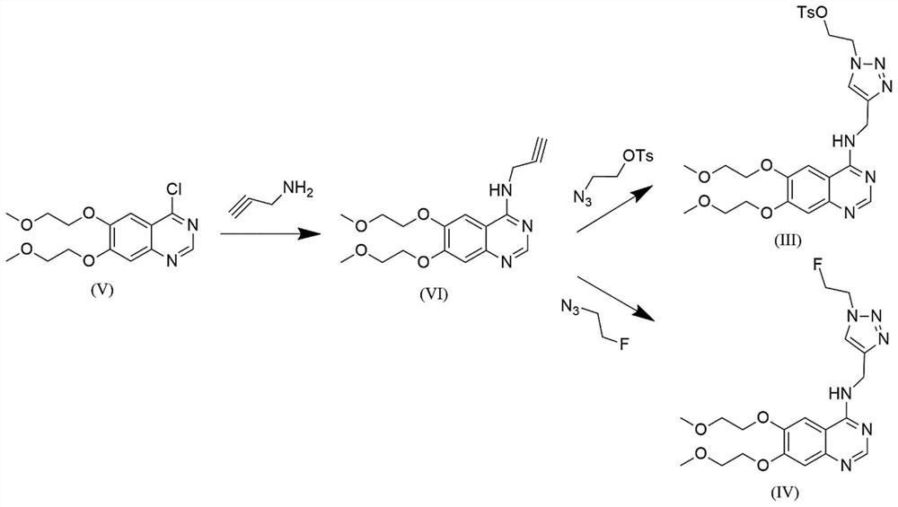
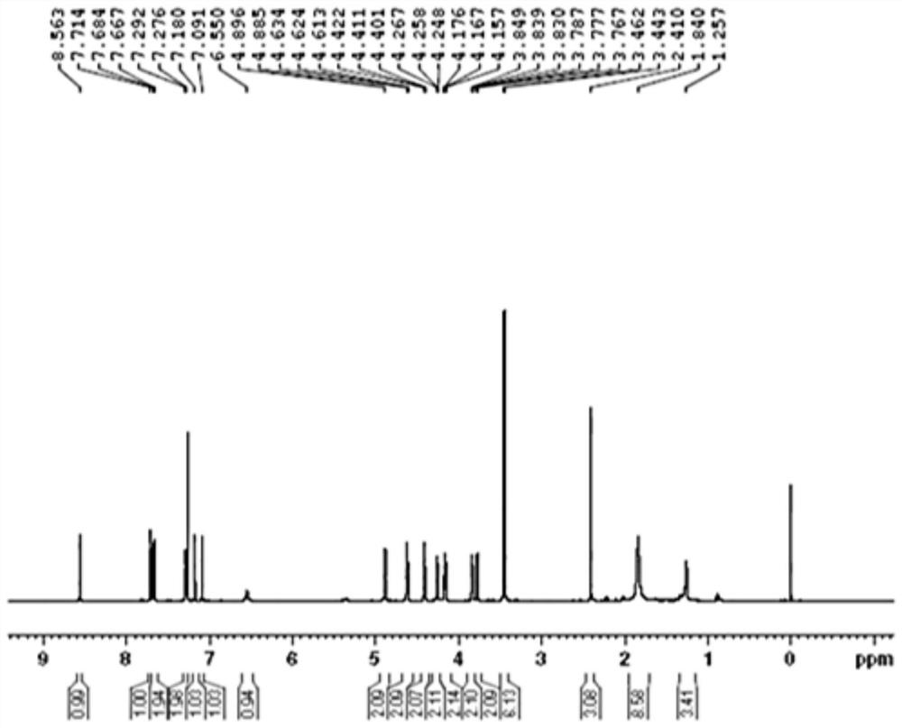
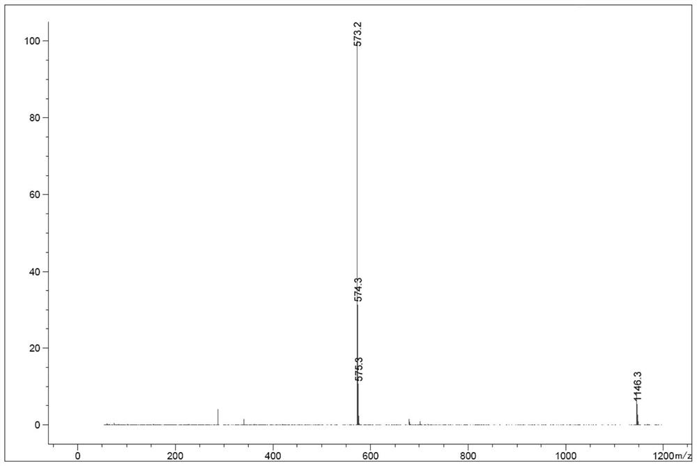
[0087] The compound represented by the formula (III) in Example 1 ( 18 Preparation method of F-labeled EGFR positron imaging agent precursor)
[0088] 4-Chloro-6,7-bis(2-methoxyethoxy)quinazoline (formula (V), 40.00g, 1eq), DMF (N,N-dimethylformaldehyde) were successively added to the 500mL three-necked flask. amide, 200 mL), potassium carbonate (35.38 g, 2 eq), propargylamine (8.48 g, 1.2 eq), stirred at 90° C. for 2.5 h. After the completion of the reaction monitored by TLC (thin layer chromatography), the temperature was lowered to room temperature (20-30° C.), 400 mL of water was added, and the mixture was stirred uniformly. Filter and wash twice with 400 mL of water. The filter cake was spin-dried to obtain 24.5 g of a silver-white solid product (formula (VI)) with a yield of 57.8%.
[0089] 4-propargylamino-6,7-bis(2-methoxyethoxy)quinazoline (formula (VI), 0.42g, 1.0 eq), copper sulfate pentahydrate (0.018g) were successively added to the reaction flask. , 0.05eq), ...
Embodiment 2
[0091] The compound represented by the formula (IV) of Example 2 ( 18 Preparation method of F-labeled EGFR positron imaging agent standard)
[0092] 4-Chloro-6,7-bis(2-methoxyethoxy)quinazoline (formula (V), 40.00g, 1eq), DMF (N,N-dimethylformaldehyde) were successively added to the 500mL three-necked flask. amide, 200 mL, 5V), potassium carbonate (35.38 g, 2 eq), propargylamine (8.48 g, 1.2 eq), stirred at 90 °C for 2.5 h. TLC (thin layer chromatography) monitoring after the completion of the reaction, the temperature was lowered to room temperature (20-30° C.), 400 mL (10 V) of water was added, and the mixture was stirred uniformly. Filter and wash twice with 400 ml of water. The filter cake was spin-dried to obtain 24.5 g of a silver-white solid product (formula (VI)) with a yield of 57.8%.
[0093] 4-propargylamino-6,7-bis(2-methoxyethoxy)quinazoline (formula (VI), 2.70g, 1.0eq), copper sulfate pentahydrate (0.40g) were successively added to the reaction flask. , 0.2eq...
Embodiment 3
[0095] The compound represented by the formula (II) in Example 3 ( 18 Preparation method of F-labeled EGFR positron imaging agent)
[0096] (1) bombarding H with a medical cyclotron 2 18 O, through 18 O(p,n) 18 F nuclear reaction produces 500mCi 18 F, and conduct it in an anion exchange column, measure the activity and use 1.5 mL of mixed solution (15.0 mg of 4,7,13,16,21,24-hexaoxo-1,10-diazabicyclo[8.8.8] 2 Hexane (K 2.2.2. ) plus 4.5mg K 2 CO 3 Dissolved in 0.15mL water and 1.35mL acetonitrile) 18 F rinsed into the reaction flask;
[0097] (2) High-purity helium gas was continuously blown into the reaction flask, water was removed azeotropically for 3 minutes at 110 °C, and dried; 5 mg of the precursor was dissolved in 1 mL of anhydrous DMSO solution, added to the reaction flask, and reacted at 110 °C for 15 min.
[0098] (3) after cooling the reaction solution, carry out semi-preparative HPLC separation to obtain 18 F-labeled EGFR positron imaging agent, the separ...
PUM
 Abstract
Description
Claims
Application Information
 - R&D
- Intellectual Property
- Life Sciences
- Materials
- Tech Scout
- Unparalleled Data Quality
- Higher Quality Content
- 60% Fewer Hallucinations
Browse by: Latest US Patents, China's latest patents, Technical Efficacy Thesaurus, Application Domain, Technology Topic, Popular Technical Reports.
© 2025 PatSnap. All rights reserved.Legal|Privacy policy|Modern Slavery Act Transparency Statement|Sitemap|About US| Contact US: help@patsnap.com


