Bioactive preparation based on tumor cell membrane, and preparation method and application thereof
A bioactive, cell membrane technology, applied in the field of medicine, to achieve the effect of inhibiting the production of tumor exosomes, improving the anti-tumor effect in vivo, and having less toxic and side effects
- Summary
- Abstract
- Description
- Claims
- Application Information
AI Technical Summary
Problems solved by technology
Method used
Image
Examples
preparation example Construction
[0060] In yet another specific embodiment of the present invention, a method for preparing the above-mentioned cell membrane-based bioactive preparation is provided, including:
[0061] The tumor cell membrane modified by sodium alginate oxide forms a gel factor, and the CDK5 inhibitor and the exosome inhibitory drug are respectively dissolved and mixed uniformly to form a drug solution, which is added to the aqueous solution of the gel factor and stirred to obtain a gelling solution.
[0062] In yet another specific embodiment of the present invention, the solvent for dissolving the CDK5 inhibitor and the exosome inhibitor is deionized water, which has a better dissolving effect, and the method of adding the drug solution is dropwise, so that the antitumor drug components are more dispersed uniform;
[0063] In yet another specific embodiment of the present invention, the cell membrane vesicles are prepared by the following method:
[0064] (1) Collect mouse tumor tissue, an...
Embodiment 1
[0095] Example 1: Preparation of tumor cell membrane vesicles modified by sodium alginate oxide
[0096] 1) Preparation of tumor single cell suspension: After collecting mouse melanoma tissue, it was obtained by adding homogenization buffer and then grinding. The homogenization buffer contained 0.25mM sucrose, 1mM EDTA, 20mM HEPES-NaOH and protease inhibitor Reagent PBS buffer.
[0097] 2) Extraction of tumor cell membrane: The collected tumor single-cell suspension was subjected to probe ultrasound at 4°C to break the tumor cells to obtain broken tumor cells. The power amplitude of the probe ultrasound was 40%, the time was 10min, and the interval was 4s. Off: 2s; Centrifuge the broken tumor cells at 3000g for 10min at 4°C to remove melanin for purification; centrifuge the purified broken cell suspension at 15000g for 60min at 4°C to obtain tumor cell membrane pellets; redisperse the cell membrane pellets In PBS, cryopreserved.
[0098] 3) Preparation of tumor cell membrane...
Embodiment 2
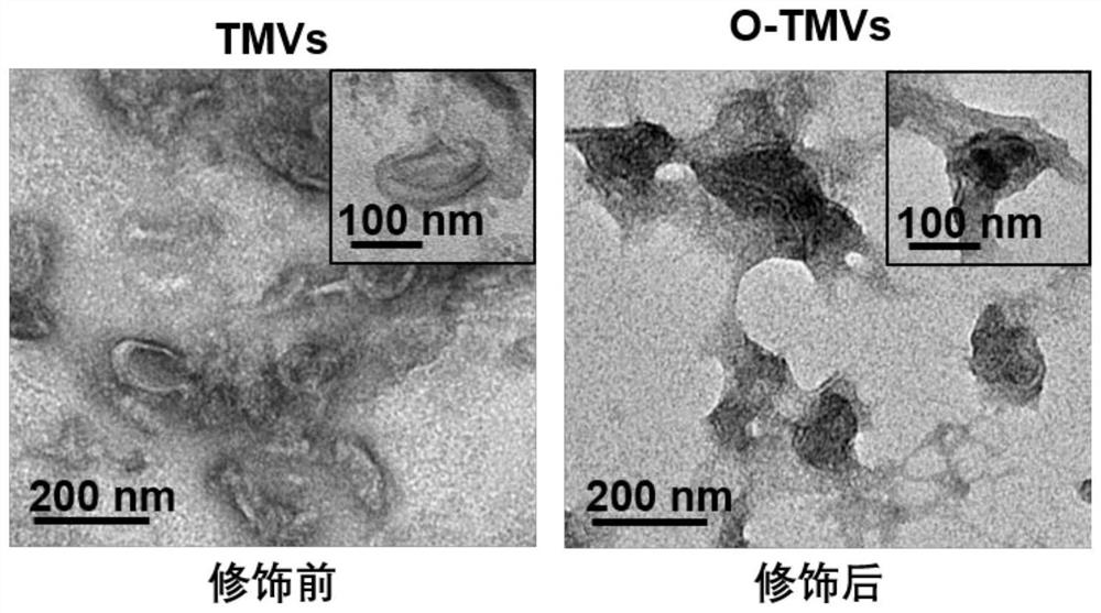
[0101] Example 2: Transmission electron microscopy (TEM) identification of nanostructures of tumor cell membrane vesicles before and after modification
[0102] Use a pipette gun to draw 10 μL of the tumor cell membrane vesicle solution before and after modification, carbon film copper grid, and filter paper to absorb excess liquid, dry it at room temperature, and observe it under a transmission electron microscope. The result is as figure 1 As shown in the transmission electron microscope pictures, it can be confirmed that the morphology of the tumor cell membrane vesicles before and after the modification is significantly different, and the sizes are ~160 and ~200nm respectively, and the size is uniform and the dispersion is good.
PUM
| Property | Measurement | Unit |
|---|---|---|
| size | aaaaa | aaaaa |
Abstract
Description
Claims
Application Information
 - R&D
- Intellectual Property
- Life Sciences
- Materials
- Tech Scout
- Unparalleled Data Quality
- Higher Quality Content
- 60% Fewer Hallucinations
Browse by: Latest US Patents, China's latest patents, Technical Efficacy Thesaurus, Application Domain, Technology Topic, Popular Technical Reports.
© 2025 PatSnap. All rights reserved.Legal|Privacy policy|Modern Slavery Act Transparency Statement|Sitemap|About US| Contact US: help@patsnap.com


