Device and method for high-purity sorting of rare cells

A rare cell, high-purity technology, applied in the field of devices for high-purity sorting of rare cells, can solve the problems of inability to apply cell sorting, low sorting speed and efficiency, complex structure and operation, etc.

Active Publication Date: 2021-09-10
ZHEJIANG UNIV
View PDF7 Cites 1 Cited by
  • Summary
  • Abstract
  • Description
  • Claims
  • Application Information

AI Technical Summary

Problems solved by technology

[0006] The optical tweezers method is currently unable to be applied to cell sorting of ...

Method used


Image

  • Device and method for high-purity sorting of rare cells
  • Device and method for high-purity sorting of rare cells
  • Device and method for high-purity sorting of rare cells

Examples


Embodiment approach

[0084] As a specific implementation, the rare cell sorting method is as follows:

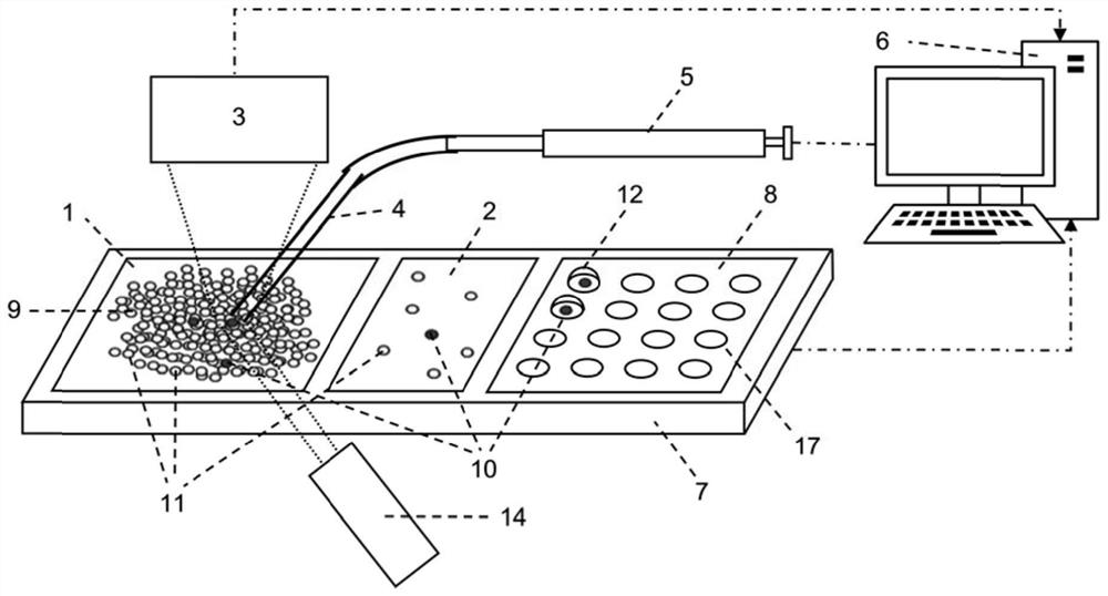
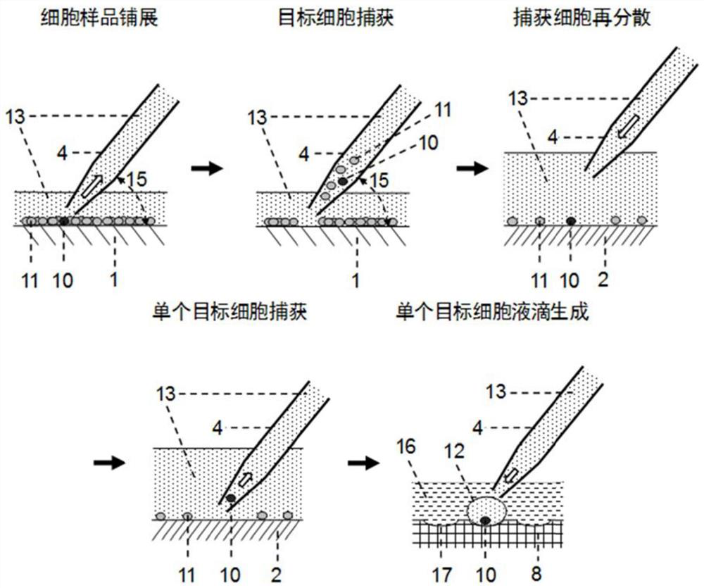
[0085] Clean flat-bottomed glass containers with ethanol, water, and EDTA in sequence (you can use figure 1 The integrated flat-bottomed container shown can also be a separately set flat-bottomed container, such as a petri dish, etc.), and the cell suspension (that is, the suspension of cell sample 9) that has been labeled with a fluorescent antibody into the flat-bottomed glass container In step 1, oscillate to disperse the cells evenly, settle naturally for 3-5 minutes, and spread the cells into a cell layer within 3 layers ( figure 2 ).

[0086] Fluorescently label target cells with specific antibodies before detection, such as Figure 4 As shown, using Cell Imaging Detector 3 (Laser Induced Fluorescence Cell Imaging Detector, such as Figure 4 ) with the use of light source 14 to scan the fluorescent signal in the detection area, and use the computer vision software based on threshold a...

Embodiment 2

[0092] In embodiment 2, device structure is as Figure 7 shown.

[0093] Pour the cell suspension containing the target cells 10 labeled with fluorescent antibodies and non-target interfering cells 11 into the culture dish 1, and incubate in the incubator for about 30 minutes. At this time, the adherent cells can form a tightly packed monolayer Adherent growth. Add an appropriate amount of trypsin cell digestion solution to digest the cells and form a low-adhesion single-cell layer at the bottom of the culture dish.

[0094] Use the motorized microscope 3 to scan the bright field and fluorescence images of the detection area, and use the image classification software based on artificial intelligence to automatically identify the fluorescent cells therein (see Example 1). Synthesize the fluorescence image recognition results under different excitation wavelengths to determine the identity of the target cells location and count its number. It is also possible to combine multi...

Embodiment 3

[0100] In embodiment 3, device structure is as Figure 9 shown.

[0101] The integrated chip 19 is used for experiments, and the integrated chip is processed with a cell spreading area (equivalent to container 1) and a multiple dispersion area (equivalent to container 2) with microstructures (including micro-pit arrays and micro-pillar arrays 18) arrays. and the area carrying the captured cell droplet (equivalent to chip 8). Pour the cell suspension containing the target cells into the cell spreading area of ​​the integrated chip, and through natural sedimentation and scraper control, a monolayer of cell spreading is formed, and the cells are bound in the micropit or the gap between the micropillars. The surface of the sample scanning area is modified in advance to inhibit the non-specific adsorption of proteins and reduce the adhesion of cells on its surface.

[0102] Use the fast and high-sensitivity cell imaging detector 3 (laser-induced fluorescence cell imaging detector...


PUM


Abstract

The invention discloses a method and device for high-purity sorting of rare cells. The method comprises the following steps of (1) spreading a cell sample; (2) carrying out scanning imaging on the spread cell sample; and (3) according to an imaging result, reading position information of target cell and distance information between the target cell and other surrounding non-target cells, and judging the obtained distance information: if the distance information does not meet a set sampling distance requirement, sampling an area where the target cell is located according to the position information of the target cell, obtaining an intermediate cell sample containing the target cell and other surrounding non-target cells, spreading the obtained intermediate cell sample again, and returning to the step (2); and if the distance information meets the set sampling distance requirement, directly sampling the target cell, so that sorting of the target cell is achieved. The method disclosed by the invention is simple and convenient to operate, and has the advantages of high cell spreading speed, high flux, high target cell sorting efficiency, high purity, low omission rate, repeatability and the like.

Description

technical field [0001] The field of the present invention is the field of rare cell sorting, in particular to a device for high-purity sorting of rare cells and its application method. Background technique [0002] Cells are the basic functional units of the morphological structure and life activities of organisms. The understanding of the laws of life activities of organisms must be based on cell research, and explore the changes of cells in the process of life activities such as growth and differentiation, metabolism and reproduction, inheritance and evolution of organisms. However, due to the randomness of the life process and the complexity of the external environment, even cells from the same source may differ greatly in terms of gene level and protein level. In recent years, a large amount of experimental evidence has shown that specific individual cell behaviors and individual differences and heterogeneity among them play an important role in many key life processes ...

Claims


Application Information

Patent Timeline
IPC IPC(8): C12N5/00C12M1/00C12M1/34C12M1/26
CPCC12N5/00C12M47/04C12M41/36C12M41/48C12M33/04C12M23/02C12N2509/10
Inventor 方群王宇王慧峰
Owner ZHEJIANG UNIV
Why Eureka
Try Eureka