Preparation method of protozoa cyst transmission electron microscope sample

A transmission electron microscope sample and protozoan technology, which is applied in the preparation, sampling, and measurement devices of test samples, can solve problems such as non-repeatability, sample preparation failure, and mixed quality, so as to promote penetration through the cyst wall and increase Effectiveness, effect of increasing permeability

Pending Publication Date: 2021-09-17
NINGBO UNIV
View PDF7 Cites 2 Cited by
  • Summary
  • Abstract
  • Description
  • Claims
  • Application Information

AI Technical Summary

Problems solved by technology

As long as one of the steps is not handled properly, it will lead to the failure of sample preparation, and the resulting internal structure will be degraded to varying degrees.
However, as far as the current experimental methods and operating techniques are concerned, the quality of the produced protozoan cyst electron microscope samples is mixed, and there are often different degrees of cavities inside the cyst, making it difficult to observe the organelle structure inside the cyst accurately and with a large field of view. It is difficult to clearly present the ultrastructure, and the failure rate is high
Moreover, there is no standard sample preparation method. The sample preparation is often based on personal experience, and the results obtained are unstable and non-repeatable.

Method used


Image

  • Preparation method of protozoa cyst transmission electron microscope sample
  • Preparation method of protozoa cyst transmission electron microscope sample
  • Preparation method of protozoa cyst transmission electron microscope sample

Examples


Embodiment 1

[0065] After optimizing the traditional method, sterilized seawater was used to dilute glutaraldehyde with a concentration of 3% as a fixative, and plant tissue embedding agent (Spurr embedding agent) was used to replace animal tissue cell embedding agent (Epon812 epoxy resin embedding agent). Embedding agent) stimulates the specific steps of Cryptocaryon cyst transmission electron microscope ultrathin section sample preparation method:

[0066] (1) Select a single Cryptocaryon cyst and the diameter should not be too large, but it is well developed, and the development of the cyst is observed under an optical microscope before fixation to see if it is the expected effect;

[0067] (2) Front fixation: Transfer the cysts selected in (1) to a corner of the glass slide, blot the excess culture medium, and use 3% glutaraldehyde (Glutaraldehyde 25% EM grade) prepared in advance to seal the cysts. Rinse the capsule into a glass fixation cylinder containing a small amount of mixed fix...

Embodiment 2

[0088] After optimizing the traditional method, a mixed solution of 3% glutaraldehyde and 0.3% Tritan X-100 was used as the fixative, and the plant tissue embedding agent (Spurr embedding agent) was used to replace the animal tissue cell embedding agent (Epon812 epoxy resin Embedding agent) stimulates Cryptocaryon cysts to the specific steps of transmission electron microscope ultrathin section sample preparation method:

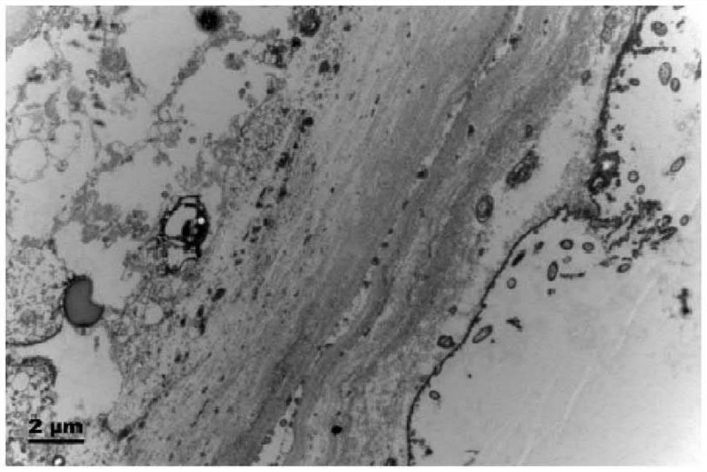
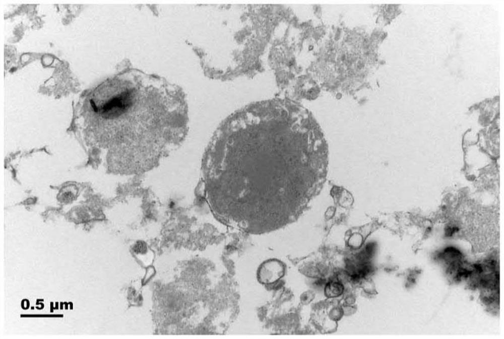
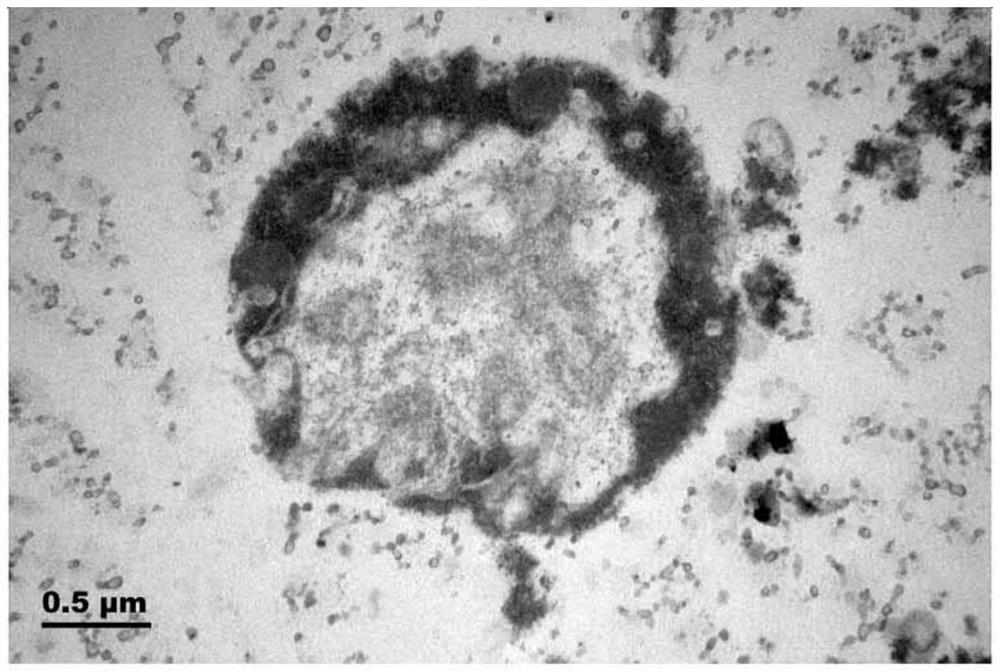
[0089] In this embodiment and embodiment 1, except using different fixatives (3% glutaraldehyde and 0.3% Tritan X-100 mixed solution), other treatments are basically the same, and the electron micrograph is as follows Figure 7-9 Shown, is to utilize the TritanX-100 mixed solution of 3% glutaraldehyde+0.3% as fixative solution, and utilizes the sample result after Spurr embedding agent processing, and traditional method ( Figure 1-3 ) compared with the electron microscope, it was found that the internal substructure of the cell is clearly identifiable, and ...


PUM

PropertyMeasurementUnit
lengthaaaaaaaaaa

Abstract

The invention discloses a preparation method of a protozoa cyst transmission electron microscope sample, which is characterized in that a traditional fixing method is improved, and glutaraldehyde and Tritan X-100 are used for treating a cyst under the condition of not damaging the cyst, the glutaraldehyde plays a fixing role, and Tritan X-100 has the characteristic of increasing the permeability of a eukaryotic cell membrane, has no killing effect on microorganisms, and can promote a stationary liquid to penetrate through a cyst wall, so that a sample is quickly fixed, and the difficulty that the sample is not fully fixed in the past is solved. An Epon812 epoxy resin embedding agent in a traditional method is replaced by the Spurr embedding agent, so that the embedding agent can better and completely permeate into the cyst. According to the method, the treatment conditions in each step are optimized according to the characteristics of the cyst, so that the effectiveness of a sample preparation result is improved, and a basis is provided for clearly presenting the sample. The method can be applied to preparation of most protozoa cyst TEM samples, and has certain stability and repeatability.

Description

technical field [0001] The invention relates to a preparation method of a transmission electron microscope sample, in particular to a preparation method of a protozoa-encapsulated transmission electron microscope sample, and belongs to the technical field of transmission electron microscope sample preparation. Background technique [0002] Protozoan cysts are mainly divided into three types: "dormant cysts", "infectious cysts" and "reproductive cysts". "Dormant cyst" is a dormant state formed by protozoa in response to an unfavorable environment, and it is common in free-living ciliates (cystophora and pseudocoronoids, etc.); "infectious cyst" is The transmission stage in the life cycle of higher animal parasites, whose chitin cyst wall has acid resistance, which can prevent the parasite body from being digested by the host (dysentery amoeba and Lampagiardia, etc.); and the above two different Yes, the cyst that undergoes normal cell division and produces larvae is a "repro...

Claims


Application Information

Patent Timeline
Patent Type & Authority Applications(China)
IPC IPC(8): G01N1/28G01N1/31G01N1/36
CPCG01N1/286G01N1/31G01N1/36G01N2001/2873
Inventor 周理耀尹飞詹萍萍孔进东谢骁
Owner NINGBO UNIV
Why Eureka
Try Eureka