Method for improving accuracy of tumor marker

A tumor marker and accuracy technology, which is applied in the field of improving the accuracy of tumor markers, can solve the problems of low markers, poor specificity, and missed opportunities for diagnosis and treatment, so as to improve accuracy, sensitivity and specificity, and enhance relative The effect of signal strength

Pending Publication Date: 2021-10-22
HENAN CANCER HOSPITAL
View PDF5 Cites 0 Cited by
  • Summary
  • Abstract
  • Description
  • Claims
  • Application Information

AI Technical Summary

Problems solved by technology

However, there are still two shortcomings that restrict its application in clinical diagnosis: First, because the signal of markers in the blood is usually very low, about 20% of tumor patients cannot detect the TM required for diagnosis in the early stage. When the clinical diagnosis value is reached, the best opportunity for diagnosis and treatment has been missed; second, the specificity of TM application is poor, and many pathological / physiological factors such as ascites, inflammation, endometriosis and other non-cancer factors can cause abnormal increase of TM , the marker signals of early tumors are buried in these noise signals and cannot be identified, and the specificity of TM application is poor. The early detection of tumors using markers must improve the sensitivity and specificity of the detection method, which hinders to a great extent. In order to measure the validity and reliability of TM in blood, it affects the detection rate of early tumors

Method used


Image

  • Method for improving accuracy of tumor marker
  • Method for improving accuracy of tumor marker
  • Method for improving accuracy of tumor marker

Examples


Embodiment 1

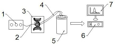
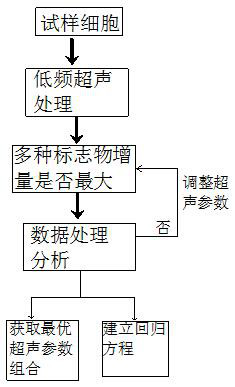
[0046] Ultrasound propagates in the medium in the form of waves, squeezes and stretches the molecular structure of the cell membrane and produces a series of microbubbles on the cell membrane. Rupture causes transient or steady-state gaps and collapses on the cell membrane, forming cavities at the corresponding positions, that is, acoustic holes. This process is called the effect produced by the cavitation mechanism of ultrasound. TM is a glycoprotein located in the cell or on the cell surface, which can be shed from the cell surface or released from the cell into the extracellular solute. Studies have shown that ultrasonic cavitation can produce 110nm holes in animal cell membranes, which is enough to allow TM to pass through. The content of TM in tumor cells is different from that in normal cells. TM passes through these holes and is released from the inside to the outside of the cell along the concentration gradient, thereby Causes the amplification of TM signal. Ultrasound...

Embodiment 2

[0058] On the basis of Example 1, when the ultrasonic signal generating component acts on the sample, microbubbles W are generated in the liquid environment of the sample. When the microbubbles are subjected to low-power ultrasonic action, steady-state cavitation occurs, resulting in A spherical force radiation is applied to the cell membrane surface of the sample to vibrate a certain area on the cell membrane surface. The vibration frequency is the same as that of the microbubble W. When the vibration frequency of the cavitation nucleus is close to the natural frequency of the cell membrane surface, due to Resonance occurs on the surface of the cell membrane, and the surface of the cell membrane is easily damaged, resulting in acoustic holes. At this time, the amplitude U of the radial velocity vector of the liquid caused by the vibration of the microbubble W is:

[0059] U=W 2 ·W min / TL 2 ; where W is the radius of the microbubble (5×10 -6 -5×10 -5 cm), L is the distanc...

Embodiment 3

[0065] To judge the degree of deterioration of the development stage of cancer, there are usually two evaluation indicators in clinical medicine: one is the stage, and the other is the degree of differentiation. The development stage of a tumor is usually determined by three numbers in the TNM system, where T represents the size of the tumor and the degree of invasion of adjacent tissues, N and M represent the degree of metastasis of local lymph nodes and distant lymph nodes, respectively . Tumor staging is a commonly used standard method to measure the degree of tumor deterioration, which is divided into stage I, stage II, stage III, stage IV and various substages. The diagnosis of tumor staging still relies on the manual diagnosis of doctors. , there is no professional computer calculation program.

[0066] The electrochemical workstation includes an algorithm module for staging the tumor markers in the sample, and the algorithm module for staging the tumor markers includes...


PUM


Abstract

The invention relates to a method for improving the accuracy of a tumor marker. The method comprises the following steps: S1, an ultrasonic signal generation assembly acts on a sample; S2, the tumor marker of the sample is captured by an electrode through a capture probe; S3, the capture probe and the electrode enrich and amplify the signal and then transmit the signal to an electrochemical workstation; S4, the electrochemical workstation judges whether the increment of the tumor marker is maximum or not, and if not, the ultrasonic signal generation assembly is controlled to adjust the ultrasonic parameters until the increment of the tumor marker is maximum; and S5, if the increment of the tumor marker is increased to the maximum, a linear relationship between the electric signal and the concentration of the sample is determined, the linear relationship is displayed through a display of an electrochemical workstation, and the sample is quantitatively detected. According to the detection system provided by the invention, the ultrasonic signal is used for amplifying the tumor marker signal, so that the relative signal intensity of the early tumor marker is enhanced, the linear relationship between the electric signal and the concentration of the standard substance to be detected is determined, and the sensitivity and specificity of early cancer detection are improved.

Description

technical field [0001] The invention belongs to the technical field of biomedical detection, and in particular relates to a method for improving the accuracy of tumor markers. Background technique [0002] The essence of cancer is the accumulation of specific genetic and epigenetic defects in DNA, resulting in abnormal gene expression. At this time, the growth mode and apoptosis pathway of cells are out of control, and such cells are cancer cells. The continuous growth of cancer cells will promote the formation of new blood vessels, which provides a channel for the metastasis of cancer cells themselves. The condition of cancer is quite complicated, including more than 200 kinds and may affect more than 60 organs of the human body. The early occurrence of cancer is basically simple , but multiple metastases to other organs are likely to occur in the middle and late stages. At this time, the treatment of cancer has reached a very difficult stage, and the cure rate is almost ze...

Claims


Application Information

Patent Timeline
Patent Type & Authority Applications(China)
IPC IPC(8): G01N27/26G01N27/30G01N27/327G01N1/28G16B40/00
CPCG01N27/26G01N27/308G01N27/3278G01N1/28G16B40/00
Inventor 李燕彭亮朱兆承付槟梵焦玥曹小琴
Owner HENAN CANCER HOSPITAL
Why Eureka
Try Eureka