Urine detection method of platinum super-nanoparticles based on hypoxia response and application of urine detection method

A detection method and ultra-nano technology, applied in the field of urine detection of platinum ultra-nanoparticles, can solve the problems of high price, low sensitivity and limited sensitivity of imaging technology.

Pending Publication Date: 2022-03-04
NANJING UNIV
View PDF0 Cites 1 Cited by
  • Summary
  • Abstract
  • Description
  • Claims
  • Application Information

AI Technical Summary

Problems solved by technology

However, imaging techniques are expensive and relatively insensitive
At present, molecular diagnosis is the main method for early diagnosis of cancer, but due to the relatively low shedding rate of biomarkers at the tumor site and the huge dilution of blood circulation, the strategy of detecting circulating endogenous biomarkers faces the inherent sensitivity of early disease limits

Method used


Image

  • Urine detection method of platinum super-nanoparticles based on hypoxia response and application of urine detection method
  • Urine detection method of platinum super-nanoparticles based on hypoxia response and application of urine detection method
  • Urine detection method of platinum super-nanoparticles based on hypoxia response and application of urine detection method

Examples


Embodiment 1

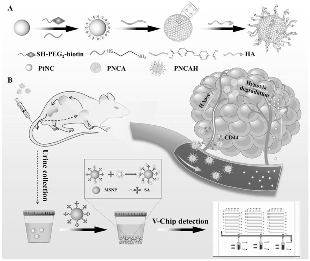
[0025] The present embodiment provides the preparation method of PNCAH nanoparticles:

[0026] PtNCs (0.01g) were treated with mercaptoethylamine (SH-NH 2 , 0.4mM) and mercaptoethanol (SH-OH, 0.6mM) for amination, the main function of mercaptoethanol is to prevent the aggregation of PtNCs, react for 3h. Then 0.002g azo was placed in 200 μL of dimethylsulfoxide (DMSO) with 0.038g N-ethyl-N'-(3-(dimethylamino)propyl)carbodiimide EDC and 0.0058g N- Hydroxysuccinimide NHS was used to activate the carboxyl group, reacted for 2 hours, and then added the activated azo solution to the aminated PtNCs under magnetic stirring. The solvent was 10mM PBS with pH 7.4, and the rotation speed was 480r. The mixture was washed 3 times with deionized water, and the collected PNCA nanoparticles were transferred to 5 mL deionized water. Then, hyaluronic acid (HA, 5 mg) was added to the PNCA solution, and reacted under 550 r magnetic stirring for 2 h, washed with centrifuged deionized water three ...

Embodiment 2

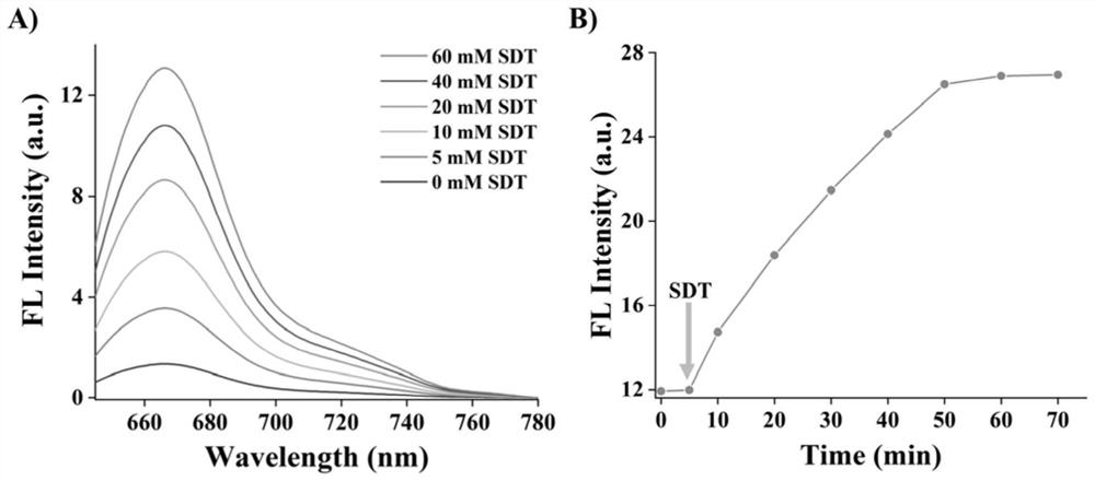
[0028] Hypoxic dissociation of PNCAH nanoparticles

[0029] This example verifies the hypoxia response of the system, the method is as follows:

[0030] First use Cy5 fluorescent dye to label PtNCs, then assemble the labeled PtNCs into PNCA nanoparticles, add hypoxia simulating agent SDT (20mM) to shake the reaction at room temperature, and measure the 670nm (Cy5 emission wavelength) every 10min. Fluorescence intensity, the result is as image 3 As shown in A, as time increases, the fluorescence intensity increases, which means that the PNCA nanoparticles dissociate; at the same time, as the concentration of SDT increases, the fluorescence intensity also increases, which also verifies the dissociation Leave( image 3 B).

Embodiment 3

[0032] Cytotoxicity of PNCAH

[0033] The cytotoxicity of the PNCAH nanoparticle of test embodiment 1, method is as follows:

[0034] Select triple-negative breast cancer cells (MDA-MB-231 cells), select different concentrations of PNCAH nanoparticles and incubate the cells, and find that the toxicity of nanoparticles to cells has no obvious toxicity below 10 μg / mL ( Figure 4 ). In order to reduce the toxic effect of PNCAH nanoparticles on cells, the concentration of PNCAH nanoparticles was controlled at 10 μg / mL in subsequent cell experiments.


PUM


Abstract

The invention discloses a urine detection method of platinum super-nanoparticles based on hypoxia response and application of the urine detection method. The platinum super-nano particle PNCA is formed by covalently connecting an aminated super-small platinum nano-cluster and 4, 4 '-dicarboxylic acid azobenzene azo with hypoxia response through an EDC/NHS reaction, and after the surface of the platinum super-nano particle PNCA is coated with a layer of hyaluronic acid HA, the nano particle can specifically target a tumor site. Afterwards, under the action of reductase in the hypoxic environment at the tumor site, the PNCA nanoparticles can be reduced and degraded into many small PtNCs to enter blood circulation again, and the PNCA nanoparticles can be excreted into urine from the kidney for disease state detection due to the fact that the particle size of the PNCA nanoparticles is smaller than the critical value 5.5 nm capable of being removed by the kidney. Due to the excellent hypoxia response effect of the PNCA nanoparticles and the efficient kidney clearing effect of the PtNCs, the PNCA/PtNCs composite material has obvious advantages in living body application.

Description

technical field [0001] The invention relates to the field of biotechnology, in particular to a urine detection method and application of hypoxia-responsive platinum supernano particles. Background technique [0002] Early diagnosis of cancer allows effective treatment of the primary tumor with local therapeutic interventions such as surgery and radiotherapy. Current strategies for early diagnosis of cancer mainly rely on imaging techniques as well as molecular diagnostic assays. Imaging techniques, such as magnetic resonance imaging (MRI) and computed tomography (CT), which can render three-dimensional images with high spatial resolution as well as high density resolution, remain the standard for clinical detection of cancer. However, imaging techniques are expensive and relatively insensitive. At present, molecular diagnosis is the main method for early diagnosis of cancer, but due to the relatively low shedding rate of biomarkers at the tumor site and the huge dilution o...

Claims


Application Information

Patent Timeline
Patent Type & Authority Applications(China)
IPC IPC(8): G01N33/48G01N31/10
CPCG01N33/48G01N31/10
Inventor 宋玉君徐琴
Owner NANJING UNIV
Why Eureka
Try Eureka