
RCAN1.4 promoter fragment for cell hypoxia and HIF1alpha protein activity indicator and application of RCAN1.4 promoter fragment

A technology of RCAN1.4 and promoter, applied in the field of genetic engineering, can solve the problems of needing four hours to overnight incubation, unstable results, high detection cost, etc., to avoid error rate and sample loss, convenient and sensitive detection, and reduce protein degradation Effect

Pending Publication Date: 2022-03-25
SHANDONG UNIV QILU HOSPITAL
View PDF0 Cites 0 Cited by
  • Summary
  • Abstract
  • Description
  • Claims
  • Application Information

AI Technical Summary

Problems solved by technology

The disadvantage of this method is that the WB operation steps are relatively cumbersome and complicated. When the cells are lysed, a sufficient amount of protease inhibitors and phosphatase inhibitors that are still within the validity period should be added to the lysate in time to slow down the degradation of the protein. Lysis, centrifugation to immunoprecipitation, etc. need to be kept at 0-4°C throughout the operation, and the reaction takes a long time, usually four hours to overnight incubation, long-term exposure to 4°C even in the presence of protease inhibitors. susceptible to protein degradation
In addition, HIF1α protein is very easy to degrade rapidly under normoxic conditions, and the operation process must be skilled and fast, otherwise the results will be affected
Furthermore, compared with other common protein antibodies of the same specification, HIF1α antibodies currently on the market have disadvantages such as high price, unstable activity, and short storage time, which have a certain impact on the accuracy of the results.
[0005] Generally speaking, this kind of method is time-consuming and laborious to detect, the result is unstable and the detection cost is high

Method used


Image

  • RCAN1.4 promoter fragment for cell hypoxia and HIF1alpha protein activity indicator and application of RCAN1.4 promoter fragment
  • RCAN1.4 promoter fragment for cell hypoxia and HIF1alpha protein activity indicator and application of RCAN1.4 promoter fragment
  • RCAN1.4 promoter fragment for cell hypoxia and HIF1alpha protein activity indicator and application of RCAN1.4 promoter fragment

Examples


Embodiment 1

[0048] Embodiment 1 plasmid construction

[0049] 1.1 Construction of RCAN1.4 promoter region RCAN1.4-315-15-LUC and RCAN1.4-315-15-GFP plasmids:

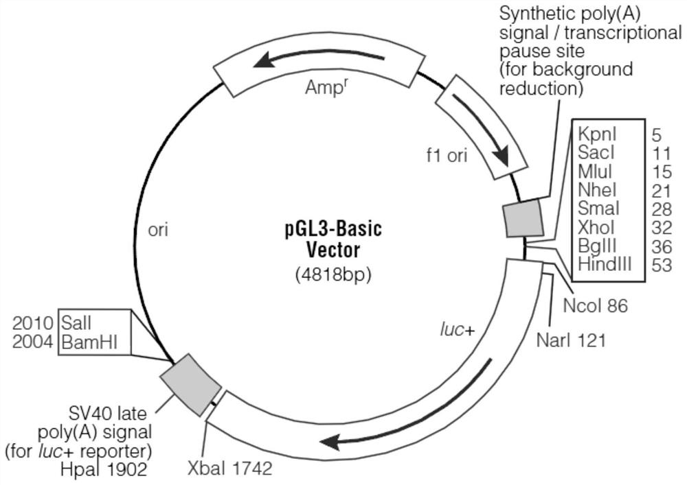
[0050] The first ATG of the first exon of RCAN1.4 was named +1, and the sequence of RCAN1.4 promoter region -315bp-15bp was inserted into the multiple cloning sites SmaI and HindIII of pGL3-basic to obtain RCAN1. 4-315-15-LUC plasmid. Under normal circumstances, the pGL3-basic vector contains the coding sequence for expressing the luciferase reporter gene LUC and has no promoter element, and cannot initiate the expression of the LUC gene. If the inserted exogenous sequence has promoter activity, it is a promoter element , then the luciferase activity of the reporter vector will increase, so the luciferase activity of the plasmid depends on the promoter activity of the insert. We excise the LUC coding sequence in the RCAN1.4-315-15-LUC plasmid and replace it with the GFP coding sequence, then when the promoter activity of the inse...

Embodiment 2

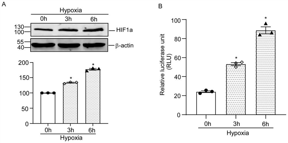
[0068] Example 2 Changes in HIF1α protein expression and luciferase activity of RCAN1.4-315-15-LUC under hypoxic conditions

[0069] 2.1 Transfect RCAN1.4-315-15-LUC into HEK293 cells using lipo3000 transfection reagent (or other brands of transfection reagents are acceptable), and the density of HEK293 cells is about 75% when transfected.

[0070] 2.2 48 hours after transfection, the cells were subjected to hypoxia (1% O 2 ) after the treatment, discard the medium immediately, wash the cells with pre-cooled PBS, add 100 μL of 1×passive lysis buffer cell lysate, place on a shaker at room temperature for lysis for 20 min until the cells are completely lysed, and transfer the cell lysate to In 1.5mL of EP, vortex shaker to mix for 15 seconds, centrifuge at 12000g for 5min at 4°C, take the supernatant and put it on ice for testing.

[0071]2.3 Using the Dual Luciferase Reporter Gene Detection Kit, add 2 μL of cell supernatant to a new EP tube in sequence, then add 10 μL of Firef...

Embodiment 3

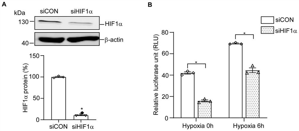
[0074] Example 3 Changes in luciferase activity of RCAN1.4-315-15-LUC after knocking down HIF1α protein under hypoxic conditions

[0075] 3.1 Use lipo3000 transfection reagent (or other brands of transfection reagents are acceptable) to co-transfect the constructed RCAN1.4-315-15-LUC plasmid with siHIF1α or siCON into HEK293 cells in a 48-well culture plate. The cell density was 75%.

[0076] 3.2 48 hours after transfection, the cells were subjected to hypoxia (1% O 2 ) after the treatment, discard the medium immediately, wash the cells with pre-cooled PBS, add 100 μL of 1×passive lysis buffer cell lysate, place on a shaker at room temperature for lysis for 20 min until the cells are completely lysed, and transfer the cell lysate to In 1.5mL of EP, vortex shaker to mix for 15 seconds, centrifuge at 12000g for 5min at 4°C, take the supernatant and put it on ice for testing.

[0077] 3.3 Using the Dual Luciferase Reporter Gene Detection Kit, add 2 μL of cell supernatant to a n...


PUM


Abstract

The invention discloses an RCAN1.4 promoter fragment for detecting a cell hypoxia state and HIF1alpha protein activity and application of the RCAN1.4 promoter fragment, and relates to the technical field of gene engineering. According to the invention, a dual-luciferase reporter gene detection system is adopted, an RCAN1.4-315-15 promoter fragment is inserted into a blank vector pGL3-basic with a firefly luciferase reporter gene, a recombinant plasmid RCAN1.4-315-15-LUC for expressing firefly luciferase is constructed, the activation condition of the RCAN1.4-315-15-LUC promoter fragment is judged by detecting the activity of the RCAN1.4-315-15-LUC luciferase, and the activity of the RCAN1.4-315-15-LUC promoter fragment is judged by detecting the activity of the firefly luciferase reporter gene. Further, the hypoxia state of the cells and the activity of the HIF1alpha protein are judged. The RCAN1.4-315-15 promoter fragment provided by the invention can be activated by the HIF1 alpha protein, has high sensitivity to the HIF1 alpha protein, and can be used as a cell hypoxia and HIF1 alpha protein activity indicator.

Description

technical field [0001] The invention belongs to the technical field of genetic engineering, and specifically relates to an RCAN1.4-315-15 promoter fragment used as an indicator of cell hypoxia and HIF1α protein activity and an application thereof. Background technique [0002] The RCAN1 gene is located in the Down Syndrome Critical Region (DSCR), including 7 exons and 6 introns, of which the first 4 exons are selective exons, and different expressions can be Four different protein isoforms are produced, the most important of which are RCAN1.1 encoded by exon 1 and RCAN1.4 encoded by exon 4. The RCAN1 gene has two transcription start sites, which are located in front of exon 1 and exon 4, and the 168 amino acids encoded by exons 5, 6, and 7 are highly conserved regions, which are expressed in all RCAN1 subtypes , and contains a calcineurin-binding domain. Studies have shown that RCAN1 has a dual regulatory effect on the signal transduction pathway of calcineurin, and partic...

Claims


Application Information

Patent Timeline
Patent Type & Authority Applications(China)
IPC IPC(8): C12N15/113C12N15/85C12N5/10C12Q1/66G01N33/58G01N33/68
CPCC07K14/47C12N15/85C12N5/0686C12Q1/66G01N33/582G01N33/68C12N2510/00C12N2830/34C12N2800/107G01N2333/47
Inventor 杨夏鑫孙秀莲运岩王频
Owner SHANDONG UNIV QILU HOSPITAL
Learn More