
Catheter stent preparation method for repairing tubular tissue and organ and apparatus thereof

A catheter stent and tissue technology, which is applied in the field of catheter stent preparation, can solve problems such as difficult control of stent pore size and microporous structure, complicated operation of fiber weaving method, and time-consuming dipping method, so as to promote tissue reconstruction and functional recovery, equipment Simple, convenient and fast effect

Inactive Publication Date: 2006-04-26
ZHEJIANG UNIV
View PDF0 Cites 25 Cited by
  • Summary
  • Abstract
  • Description
  • Claims
  • Application Information

AI Technical Summary

Problems solved by technology

Among them, the dip coating method is time-consuming and cumbersome to operate; the fiber weaving method is complicated to operate, and it is difficult to control the pore size and microporous structure of the scaffold.

Method used


Image

  • Catheter stent preparation method for repairing tubular tissue and organ and apparatus thereof
  • Catheter stent preparation method for repairing tubular tissue and organ and apparatus thereof
  • Catheter stent preparation method for repairing tubular tissue and organ and apparatus thereof

Examples


Embodiment 1

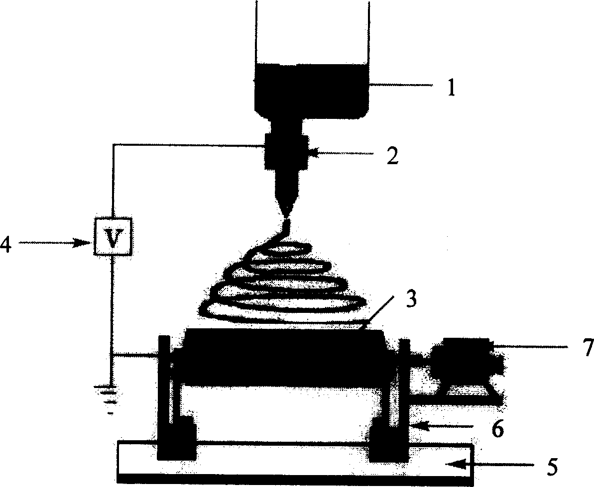
[0024] Polycaprolactone (PCL, Mn 42,500, Aldrich) 2g was dissolved in 10mL dimethylformamide (DMF) / chloroform mixed solvent (volume ratio 4 / 6), placed figure 1 In the feeder shown, the feeding speed of the spinning solution is controlled by a syringe pump; the spinning voltage is 10kV; a mandrel with a diameter of 6mm is selected, the mandrel speed is 200 rpm, and the axial speed is 0-0.15 m / s . According to the above conditions, by changing the collection time, tubular fiber scaffolds with an inner diameter of about 6 mm and different wall thicknesses can be obtained. The fiber diameter is around 750nm.

Embodiment 2

[0026] Polycaprolactone (PCL, Mn 42,500, Aldrich) 2g was dissolved in 10mL dimethylformamide (DMF) / chloroform mixed solvent (volume ratio 4 / 6), placed figure 1 In the feeder shown, the feeding speed of the spinning solution is controlled by a syringe pump; the spinning voltage is 10kV; a mandrel with a diameter of 1mm is selected, the mandrel speed is 200 rpm, and the axial speed is 0-0.15 m / s . According to the above conditions, by changing the collection time, tubular fiber scaffolds with an inner diameter of about 1 mm and different wall thicknesses can be obtained. The fiber diameter is around 700nm.

Embodiment 3

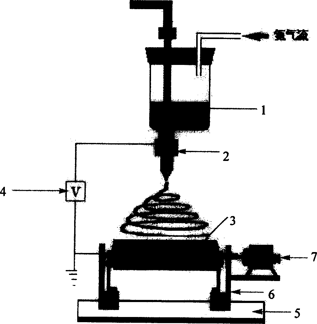
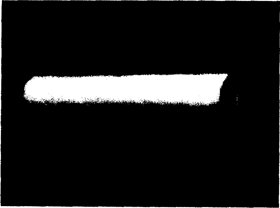
[0028] Polycaprolactone (PCL, Mn 42,500, Aldrich) 3g was dissolved in 10ml dimethylformamide (DMF) / chloroform mixed solvent (volume ratio 3 / 7), placed figure 1 In the outer chamber of the feeder shown; 2g dextran (Mn70,000, domestic) and 400mg BSA were dissolved in 10ml deionized water, placed in figure 1 In the feeder shown in the inner chamber; the PCL solution in the outer chamber is extruded from the spinning head by nitrogen flow, and the dextran solution in the inner chamber is pushed out of the spinning head by a syringe pump, and the applied voltage is 14.0kV; the diameter is 1.5mm Mandrel, the mandrel rotating speed is 300 rpm, and the axial speed is 0-0.2 m / s. Under the above conditions, a tubular scaffold composed of core-shell nanofibers with PCL as the shell and dextran / BSA as the core can be conveniently prepared, with a fiber diameter of about 600 nm. image 3 Optical photographs for the preparation of large-diameter catheter stents. Figure 4 is a scanning el...


PUM

PropertyMeasurementUnit
diameteraaaaaaaaaa
diameteraaaaaaaaaa
diameteraaaaaaaaaa

Abstract

The invention discloses a method for preparing the tube bracket restoring the tubular tissues and organs and its device. The oil soluble and water-soluble medicine or their mixture with two identical or different biological polymer solution through the single layer, inner and outer layer or compartment spinneret form the nanometer or micro fiber under the action of high voltage and static electricity, the fiber accumulates on the core rod to form the tube bracket restoring the tubular tissues and organs. The invention is characterized by the simple equipment, convenient operation and little energy consumption; the prepared tube bracket possesses larger degree of porosity; tubes of different inner diameter and thickness can be prepared by changing the electrical weaving to meet different needs; the medicine can be put into the tube at the time preparing the tubular bracket and can be released slowly in the tube. The tube bracket prepared in this invention can be used for the restoration of tubular tissure such as vessel, nerve, esophagus, tracheal and ureter.

Description

technical field [0001] The invention relates to a method for preparing a catheter bracket for repairing tubular tissues and organs and a device thereof. Background technique [0002] Tissue engineering is based on the principles of engineering and life sciences, using normal tissue cells with specific biological activity combined with biological materials to construct tissues, organs or their biological substitutes in vitro or in vivo to maintain, repair, regenerate or The science of improving the function of damaged tissues and organs. Because of its far-reaching significance in the field of scientific research and clinical application, it has received more and more attention. At present, there have been many reports on artificial skin, artificial bone tissue, and artificial blood vessels. The tubular stent prepared by the electrospinning method of the present invention is mainly suitable for the repair of tubular tissues and organs, such as blood vessels, nerves, esophag...

Claims


Application Information

Patent Timeline
Patent Type & Authority Applications(China)
IPC IPC(8): A61L29/00A61F2/00A61L27/00
Inventor 蒋宏亮赵鹏程朱康杰
Owner ZHEJIANG UNIV
Learn More