Method for in vitro separation of full-thickness retina tissue

A technology of retina and tissue slices, applied in biochemical equipment and methods, tissue culture, prosthesis, etc., can solve the problems of lack of retina and achieve good tissue activity

Inactive Publication Date: 2007-05-16
THE FIRST AFFILIATED HOSPITAL OF THIRD MILITARY MEDICAL UNIVERSITY OF PLA
View PDF1 Cites 4 Cited by
  • Summary
  • Abstract
  • Description
  • Claims
  • Application Information

AI Technical Summary

Problems solved by technology

In 2000, US patent specification US-6045791 disclosed a method for retinal pigment epithelial cell transplantation, which adopted the method of culturing the retinal pigment epithelial cells on the collagen layer in vitro, and inhaling the retinal pigment epithelial cells through the transplantation tube, but in this way only the retinal pigment epithelial cells were transplanted. Pigment epithelium, the other 9 layers of cells that lack the retina
However, the use of precise excimer laser ablation of the choroid to obtain a full-thickness retinal tissue sheet with a well-structured layer has not yet been reported.

Method used


Image

  • Method for in vitro separation of full-thickness retina tissue
  • Method for in vitro separation of full-thickness retina tissue
  • Method for in vitro separation of full-thickness retina tissue

Examples


Embodiment 1

[0043] The preparation of embodiment 1 gelatin sheet

[0044] Weigh 2.5 grams of gelatin powder, put it into a sterilized small empty bottle, add 5 milliliters of sterile normal saline to make a concentration of 50%. After inserting a needle on the bottle cap, put it into a water bath and heat to boil. After the gelatin particles are completely melted, cool slightly and pour into a cylindrical mold with a diameter of 7mm. After cooling completely, remove the gelatin from the mold, cut it into a round gelatin sheet with a thickness of 150 microns with a vibrating slicer, and immerse it in 4°C normal saline for later use. .

Embodiment 2

[0045] Example 2 Preservation of full-thickness retinal slices

[0046] Take out the donor's full-thickness retina with choroid, spread it quickly on the surface of the gelatin sheet, with the choroid facing up, and immerse it in the preservation solution within 4 hours. The preservation solution can be commercially available Ames' solution, mid-term preservation solution, frozen agent, and No. 1 antifreeze and No. 2 antifreeze. (1) The composition of Ames' solution is: NaCl 120 (mM / L), KCl3.6 (mM / L), MgSO 4 1.2 (mM / L), CaCl 2 1.2 (mM / L), NaHCO 3 23 (mM / L), NaH 2 PO4 0.1(mM / L), Na 2 HPO4 0.4 (mM / L), Glucose 10 (mM / L). (2) The composition of the medium-term preservation solution is: take the cell culture medium DMEM as the base solution, add 0.05mmol / L dexamethasone (Shanghai Xinhua Pharmaceutical Factory), 2.5% chondroitin sulfate (US Sigma Company), 1% dextran (molecular weight 4×10 4 , Shanghai Chemical Reagent Factory), 10 5 U / L tobramycin, 0.03% L-glutamine, adj...

Embodiment 3

[0049] Embodiment 3 Laser micromachining and layer removal processing of full-thickness retinal sheet

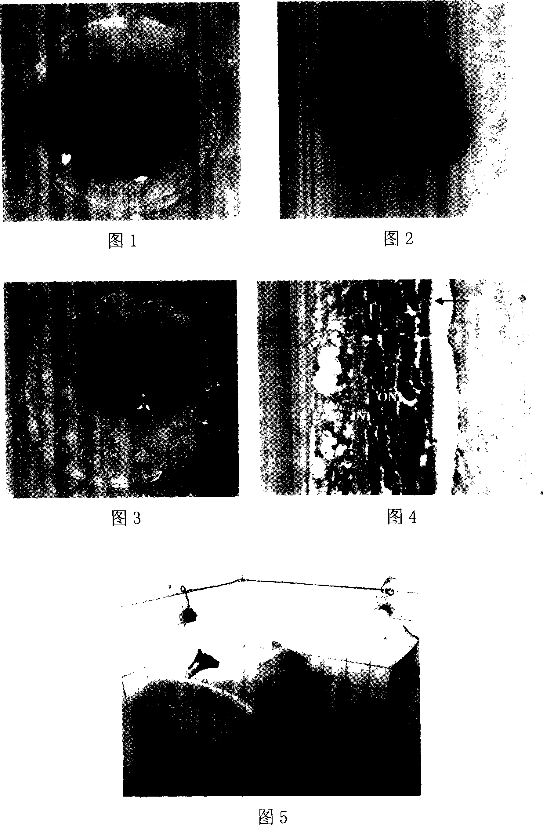
[0050] The full-thickness retinal slice with the choroid attached was laid flat on the surface of the gelatin sheet with the choroid side up (Figure 1). Absorb the residual liquid on the surface of the choroid, using the 193nm excimer laser machine of American Eagle Eye System, with a pulse frequency of 400Hz, a tracking frequency of 400Hz, a laser spot of 0.95mm, a pulse energy of 1.6mJ, and a delay time of 4-8ms. For spot scanning, the aiming light is focused on the center of the anterior surface of the choroid, and the laser is emitted in the optical keratomileusis mode (Figure 2) until the white color of the cut area can be seen under the naked eye (Figure 3). The cutting depth of optical keratomileusis mode is 50-85 microns. The tissue after laser microcutting the choroid was double-sided embedded in gelatin, melted in a 37-degree water bath for 2 minutes to make a ful...


PUM


Abstract

The invention relates to a method for externally separated preparing retina organism sheath, wherein it comprises 1, preparing gelatin sheet that adding germ-free physiological salt water into gelatin powder, heating, boiling and fusing, cooling in cylinder module, using vibration cutter to cut it into gelation sheets; 2, storing the retina organism sheet that laying the retina adhered with choroids on the surface of gelation sheet while the choroids is upward, to be stored in storage liquid; 3, laser micro cutting the retina sheet that using quasi-molecular laser, using the optical cornea embed mode to emit laser, to cut off the choroids, then packing it with gelation, emerging it in storage liquid. The invention can separate the retina organism from choroids, with integral structure and organism activity.

Description

technical field [0001] The invention relates to a method for taking full-thickness retinal tissue from a donor, separating it in vitro, and preparing it into a graft. technical background [0002] Since Royo first reported transplanting the retina into the anterior chamber of rats in 1959, researchers have found that the subretinal space between the retinal neuroepithelium and the pigment epithelium and the anterior chamber are relatively immune-privileged areas, and the graft is less affected. immune rejection. So people successively carried out retinal pigment epithelial cells, retinal photoreceptor cells, embryonic stem cells, neural stem cells, bone marrow mesenchymal stem cells, retinal stem cells, retinal neuroepithelium, full-thickness retina (retinal pigment epithelium + retinal neuroepithelium) and retina chips, Transplantation of retinal prostheses into the subretinal space of host animals (recipients). Especially in the past 20 years, researchers have used healt...

Claims


Application Information

Patent Timeline
Patent Type & Authority Applications(China)
IPC IPC(8): A61F2/14A61F9/007A61N1/02C12N5/06C12N5/08C12N5/02C12N11/00A61L27/00C12N5/071
Inventor 阴正勤陈少军李世迎余涛刘勇
Owner THE FIRST AFFILIATED HOSPITAL OF THIRD MILITARY MEDICAL UNIVERSITY OF PLA
Why Eureka
Try Eureka