Ethylsulfonated hyaluronic acid biopolymers and methods of use thereof

a technology of hyaluronic acid and biopolymer, which is applied in the direction of biochemistry apparatus and processes, unknown materials, and capsule delivery, etc., can solve the problems of limited use of hya in medical applications, limited potential use of sulfated gag biopolymers, and limited current use of hya

Inactive Publication Date: 2018-06-19
RGT UNIV OF CALIFORNIA
View PDF9 Cites 0 Cited by
  • Summary
  • Abstract
  • Description
  • Claims
  • Application Information

AI Technical Summary

Benefits of technology

[0002]Hyaluronic acid (HyA) is a natural glycosaminoglycan (GAG) biopolymer with a variety of favorable biological properties, such as promoting cell growth, inhibiting thrombosis and modulating the tissue distribution of secreted growth factors. It is currently used in a variety of products to improve surface biocompatibility, as a substrate for tissue engineering and to control drug pharmacodynamics. A significant limiting factor for the use of HyA in medical applications is its degradation in vivo due to the high endogenous concentration of hyaluronidase and other glycolytic proteins. Depending on the tissue, the half-life of HyA can be as short as minutes, and when in contact with the blood, its half-life is approximately 1.5 hours. Therefore, current uses of HyA are limited to applications where its biomaterial properties are only required for a short duration of time.
[0004]To improve the stability of HyA in vivo there are several methods of synthetic GAG sulfation. However, the primary drawback to current sulfation methods is that the chemical reactions must be performed in organic solvents that significantly complicate the process of translating the final product into a biocompatible formulation. Furthermore, a high degree of sulfation (substitution ratios of 2-4 sulfates per disaccharide unit) is required to satisfactorily resist enzymatic degradation. Thus, extensive modification of the HyA molecule limits the ability to perform additional chemical reactions on the biopolymer to improve its utility.

Problems solved by technology

A significant limiting factor for the use of HyA in medical applications is its degradation in vivo due to the high endogenous concentration of hyaluronidase and other glycolytic proteins.
Therefore, current uses of HyA are limited to applications where its biomaterial properties are only required for a short duration of time.
The potential uses for the sulfated GAG biopolymers are therefore limited in comparison to HyA, which is routinely synthesized with molecular weights exceeding 1.0 MDa.
However, the primary drawback to current sulfation methods is that the chemical reactions must be performed in organic solvents that significantly complicate the process of translating the final product into a biocompatible formulation.
Thus, extensive modification of the HyA molecule limits the ability to perform additional chemical reactions on the biopolymer to improve its utility.

Method used


Image

  • Ethylsulfonated hyaluronic acid biopolymers and methods of use thereof
  • Ethylsulfonated hyaluronic acid biopolymers and methods of use thereof
  • Ethylsulfonated hyaluronic acid biopolymers and methods of use thereof

Examples


example 1

on of Hyaluronic Acid

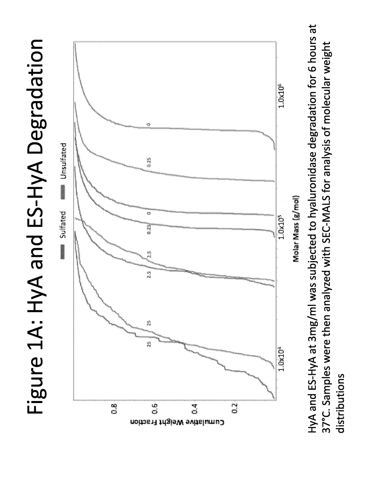
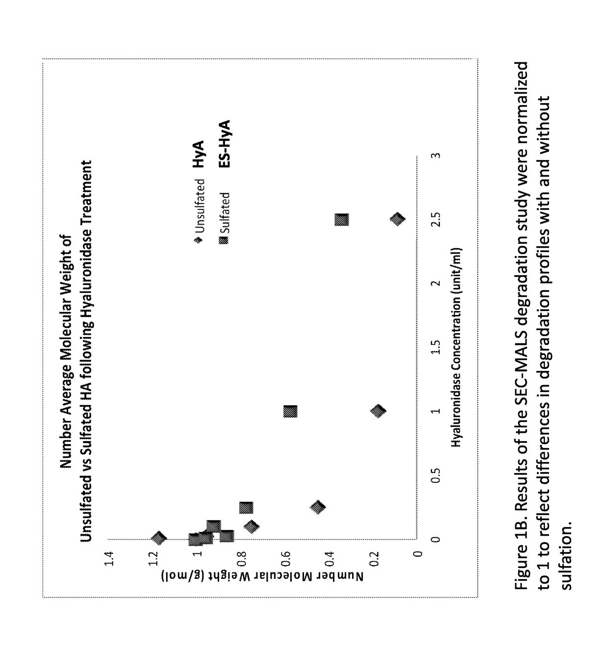
[0135]ES-HyA was synthesized using via conjugation of the aminoethylsulfonate to the HyA backbone. The ability of ES-HyA to resist enzymatic degradation relative to HyA was evaluated by incubating ES-HyA and HyA with hyaluronidase at various concentrations for 2.5 hours (FIG. 1). The cumulative molecular weight distribution was evaluated at various time points during the experimental interval using size exclusion chromatography with multi-angle light scattering analysis (SEC-MALS). It was found that the ES-HyA was able to resist the activity of the glycolytic enzyme, exhibited by molecular weights that were 2-3 times greater than the HyA at physiologically relavant concentrations of hyaluronidase (0.25-2.5 units / mL). At these concentrations, the half-life of ES-HyA appeared to be in the range of 6.6-26.9 hrs, whereas the half-life of HyA was in the range of 2.35-7.5. Thus the method of sulfonation increased the half-life of the biopolymer by approximately 3× on ...

example 2

of Sulfated Hyaluronic Acid

[0138]Sulfated hyaluronic acid was synthesized by a two-step process. First, aldehyde derivative of hyaluronic acid (HyAALD) was synthesized by sodium periodate oxidation of hyaluronic acid (HyA). HyA of 1 million Daltons was incubated with sodium periodate (NaIO4) in the dark at room temperature to convert the vicinal diols to adjacent aldehydes to produce HyAALD (FIG. 4A). Second, sulfated hyaluronic acid (SHyA) was synthesized by conjugation of 2-aminoethyl hydrogen sulfate to HyAALD and subsequent reduction using sodium cynoborohydride. HyAALD produced in the first reaction step was combined with 2-aminoethyl hydrogen sulfate (NHCH2CH2—SO4−) and the reaction product was incubated with sodium cynoborohydride (NaBH3CN) at room temperature to produce SHyA (FIG. 4B).

[0139]Validation of sulfation of hyaluronic acid was performed by NMR analysis. Proton-(1H)-NMR spectra were produced and compared for HyAALD and SHyA to demonstrate the disappearance of aldehy...

example 3

f HyA Sulfation on Cells

[0145]The viability, proliferation, and adhesion of cardiac progenitor cells (CPC) was assessed in sulfated hyaluronic acid (SHyA) with varied degrees of sulfation. Degree of sulfation was varied by varying the moles of 2-aminoethyl hydrogen sulfate reacted with HyAALD (25, 50, or 100 moles of 2-aminoethyl hydrogen sulfate). Cells encapsulated in sulfated HyA hydrogels demonstrated high viability after one day in culture, as assessed by double staining with calcein (green, live cells) and propidium iodide (red, dead cells) (FIG. 11, top panels). CPCs were capable of adhering and spreading within the sulfated hydrogel networks containing the adhesive ligand bspRGD(15), as assessed by double staining for f-actin stress fibers (TRITC-phalloidin, red) and nuclei (DAPI, blue) (FIG. 11, bottom panels).

[0146]CPC differentiation was also assessed in sulfated hyaluronic acid (SHyA) with varied degrees of sulfation. CPCs encapsulated in sulfated hydrogels containing bs...


PUM

PropertyMeasurementUnit
molecular weightaaaaaaaaaa

Abstract

The present disclosure provides methods for sulfonation of hyaluronic acid. The present disclosure provides sulfonated hyaluronic acid, and compositions, including pharmaceutical compositions, comprising the sulfonated hyaluronic acid. The present disclosure provides implantable materials and drug delivery compositions comprising a subject sulfonated hyaluronic acid.

Description

CROSS-REFERENCE[0001]This application claims the benefit of U.S. Provisional Patent Application No. 61 / 807,660, filed Apr. 2, 2013, which application is incorporated herein by reference in its entirety.INTRODUCTION[0002]Hyaluronic acid (HyA) is a natural glycosaminoglycan (GAG) biopolymer with a variety of favorable biological properties, such as promoting cell growth, inhibiting thrombosis and modulating the tissue distribution of secreted growth factors. It is currently used in a variety of products to improve surface biocompatibility, as a substrate for tissue engineering and to control drug pharmacodynamics. A significant limiting factor for the use of HyA in medical applications is its degradation in vivo due to the high endogenous concentration of hyaluronidase and other glycolytic proteins. Depending on the tissue, the half-life of HyA can be as short as minutes, and when in contact with the blood, its half-life is approximately 1.5 hours. Therefore, current uses of HyA are l...

Claims


Application Information

Patent Timeline
Patent Type & Authority Patents(United States)
IPC IPC(8): C08B37/08A61K35/545A61K35/36A61K35/34A61K35/30C08L5/08A61K9/48
CPCC08B37/0072A61K9/4816A61K35/30A61K35/34A61K35/36A61K35/545C08L5/08C12N2533/80
Inventor JHA, AMIT K.ALTIOK, EDA ISILJACKSON, WESLEY M.HEALY, KEVIN E.
Owner RGT UNIV OF CALIFORNIA
Why Eureka
Try Eureka