
Method and apparatus for non-invasive real-time biomedical imaging of neural and vascular activity

a biomedical imaging and real-time technology, applied in the field of medical imaging, can solve the problems of microwave imaging, inability to produce sharp images of deep structures within the human head or body, and invention that fails to provide real-time spatial measurements of brain functional operations

Active Publication Date: 2020-05-26
FURAXA
View PDF38 Cites 8 Cited by
  • Summary
  • Abstract
  • Description
  • Claims
  • Application Information

AI Technical Summary

Benefits of technology

The patent describes a system that uses microwave pulses to collect data from the brain. The pulses are transmitted through the skull and reflected off of its structures. The system measures the timing and amplitude of the received energy, and uses this information to map the structural features of the brain. This allows for non-invasive, whole brain imaging in a fraction of a second. The system can also be used to detect and map changes in brain tissue due to metabolic or functional changes. This has applications in brain computer interfaces and non-invasive external control of limbs, as well as identifying thought patterns and creating a general purpose mental user interface.

Problems solved by technology

Microwave imaging, however, prior to the present invention has failed to provide real-time spatial measurements of functional operation in the brain.
To date, none has produced sharp images of deep structures within the human head or body.
Also, these instruments are not sufficiently sensitive and do not have adequate signal collection efficiency to reliably detect, in real time, reflectivity or transmissivity changes in tissues that are due to functional changes due to neuronal, metabolic, or vascular changes, such as what would be measured by MEG, fMRI, fNIR, SPECT or PET.
These limitations of swept frequency microwave imaging systems are in part due to insufficient signal to noise ratio (SNR), due to the prohibitive expense of using a sufficiently large number of concurrently active TDR or Vector Network Analyzer channels and the sheer size of the large bundles of thick microwave cabling that would be required to operate concurrently to achieve acceptable signal collection efficiency.
Additionally the SNR of VNAs is insufficient to achieve acceptable signal collection efficiency in the time frame required to accurately detect real-time neurally-modulated dielectric and mechanical changes in brain tissue.
Prior art microwave imaging system using VNAs lack the sensitivity needed to detect these tiny changes, and hence, functional brain measurements using microwaves have not been usable for temporal studies of brain function nor for thought identification or control of prosthetic devices.
While prior art microwave imaging systems that use electrical pulses and sample apertures instead of swept sine waves produce potentially better images, to date these images have only been made for shallow structures in the body, such as for detecting breast tumors.
MRI, including fMRI, while enabling high resolution functional and structural images of the brain, and able to show functional and vascular activity, is non-portable and expensive.
Additionally, the 1.5 to 4 second response time that is characteristic of fMRI is too slow to display millisecond-scale real-time neural activity.
While such absorbed energy has not been shown to be harmful, long term safety also has not been proven, and it would be desirable to have no more than milliwatts of RF power transmitted into the body.
Finally, fMRI requires the subject to be confined within a large MRI machine, and remain still, and therefore cannot be used to study neural or vascular function in many operational environments, such as when performing physical tasks or job functions.
PET and SPECT both require expensive, non-portable scanners, and injection of radioactive tracers into the body.
Both methods are also very slow, and therefore unusable for real-time studies of brain activity. fNIR and EROS imagers are semi-portable, and capable of near-real-time assessment of metabolic activity in the brain, but suffer from low resolution and a poor penetration depth of approximately 1 cm.
EEG is capable of showing the summation of the real-time neural activity in a given region of the brain, but suffers from the inability to localize the source of said activity, particularly when concerning deep structures within the brain, and therefore is not considered a true imaging modality.
MEG is capable of real-time localization of neural activity in the brain, but requires a large expensive machine using superconductive detectors, and therefore is not usable in operational environments.
In addition to the attendant patient risk due to the required surgeries, the electrodes eventually become non-functional, and ECoG only provides good source localization for regions near the electrode locations.

Method used


Image

  • Method and apparatus for non-invasive real-time biomedical imaging of neural and vascular activity
  • Method and apparatus for non-invasive real-time biomedical imaging of neural and vascular activity
  • Method and apparatus for non-invasive real-time biomedical imaging of neural and vascular activity

Examples


Embodiment Construction

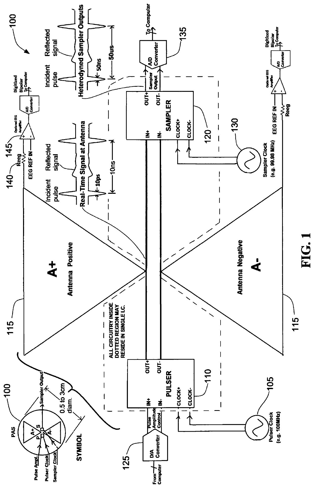
[0037]The present invention provides a compact, sensitive, and low cost waveform-edge-based medical imaging system using one or more assemblies each containing an electrical pulse or square wave generator, an antenna or microwave probe, and an electrical sampler. The pulse generator, herein also termed a pulser, is located on an electrical circuit and generates an electrical pulse, meaning a short burst, of near-Gaussian shape in some embodiments. Pulses are fed to the antenna that in turn emits an electromagnetic wave whose amplitude envelope is essentially that of the electrical pulse's shape. In use, the antenna is oriented so the electromagnetic wave propagates into the surface of the body over a region of interest. The antenna also receives energy reflected from the body in response to its own pulses, and can further receive energy from pulses which propagated through the body from one or more other pulsers with their antennae. An electrical sampler, optionally located within t...


PUM


Abstract

Biomedical images of both anatomical structure and real-time changes in neuronal, metabolic, positional, and vascular function in humans and animals is described. Ultra-wideband (UWB) pulse or square wave generators and electrical samplers, implemented using integrated circuits are used to make arrays of miniaturized microwave modules that are placed around the portion of interest in the body or head, allowing images to be made through either time-domain transmission of these pulsed waves through the body, or time domain reflectivity of the waves from internal structures, or their combination. Signal processing separate and extract the time-varying functional information from the static structural image data. The time-varying functional information from certain brain regions can be interpreted in order to control prosthetics, Brain-Machine-Interfaces and the like.

Description

BACKGROUNDField of the Invention[0001]The present invention is in the field of medical imaging and more particularly is directed to functional and structural medical imaging based on the transmission and reflection of short radio-frequency pulses, especially as applied to the brain, and applications thereof to medical testing as well as to user interfaces based on imaged brain activity.Related Art[0002]It is known that the diverse dielectric properties of biological tissues can be used to produce structural images of biological systems, such as the human body. It is also known that changes in dielectric properties of brain tissues may be measured continuously, and are indicative of changing concentrations of chemical substances, such as glucose, oxyhemoglobin and deoxyhemoglobin in various tissue regions. Additionally, it is known that when a neuron is excited it briefly expands, as do associated glial cells, such as astrocytes. When large groups of neurons in a region concurrently ...

Claims


Application Information

Patent Timeline
IPC IPC(8): A61B5/021A61B5/0476A61B5/0456A61B5/352
CPCA61B5/021A61B5/0456A61B5/0476A61B5/0507A61B2576/026A61B2562/046A61B2562/0228A61B5/0042A61B5/7225A61B5/14532A61B5/291A61B5/352A61B5/369
Inventor LIBOVE, JOELINGLE, MIKESCHRIEBMAN, DAVID
Owner FURAXA
Learn More