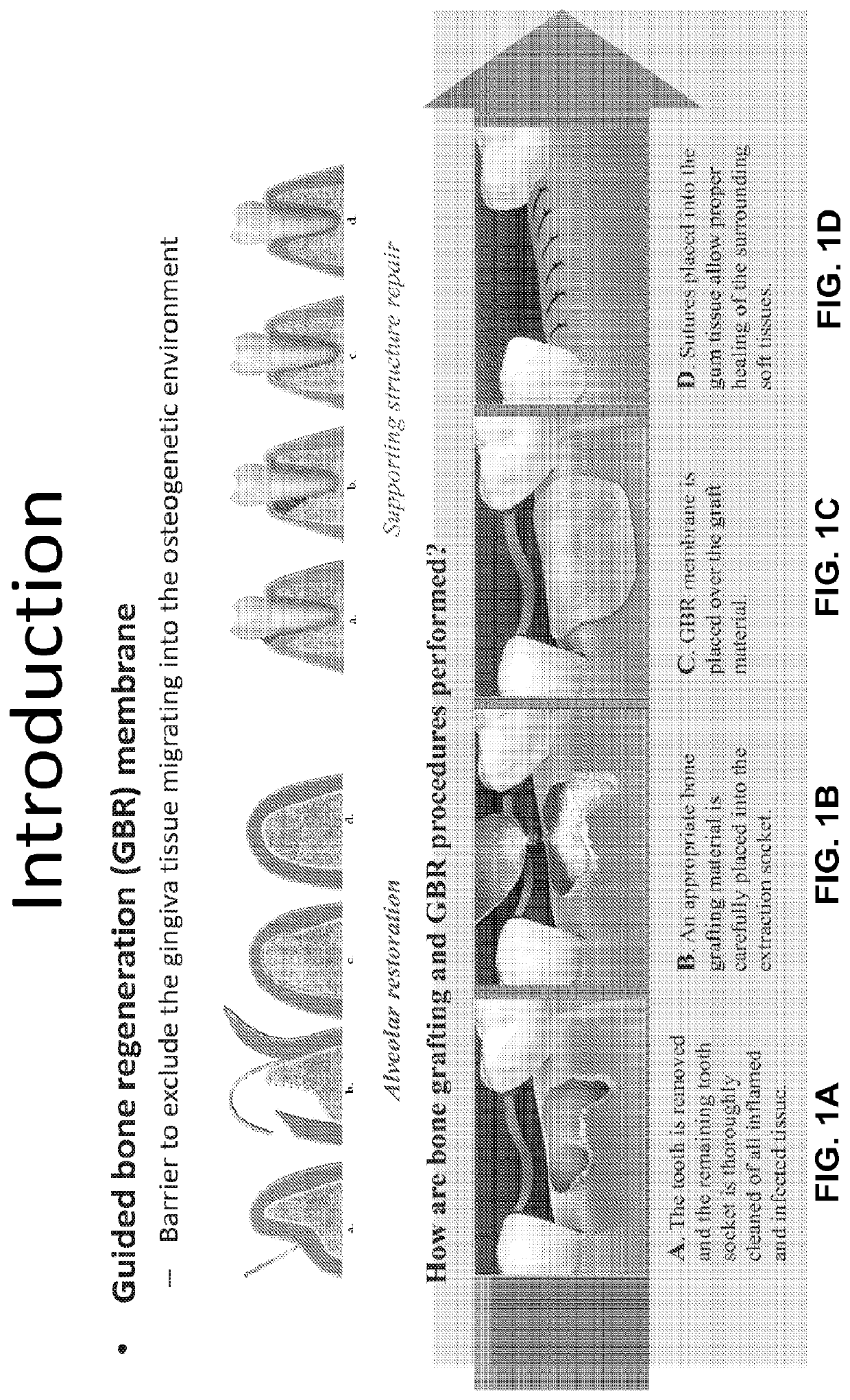
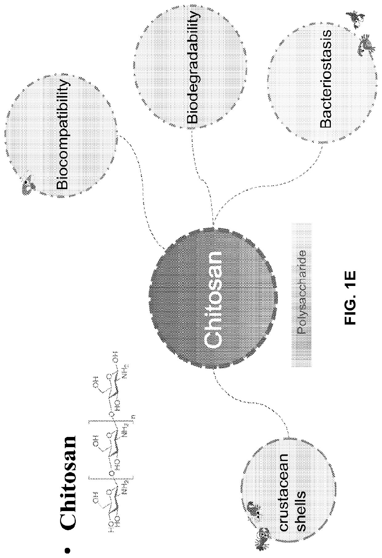
Chitosan nanofiber compositions, compositions comprising modified chitosan, and methods of use
a technology of chitosan nanofibers and compositions, which is applied in the direction of monocomponent protein artificial filaments, prosthesis, bandages, etc., can solve the problems of affecting the healing affecting the healing effect of the graft area, and the burden on both patient and physician, so as to achieve minimal swelling, minimal loss of chitosan nanofiber structure, and minimal dissolution of chitosan nanofibers
- Summary
- Abstract
- Description
- Claims
- Application Information
AI Technical Summary
Benefits of technology
Problems solved by technology
Method used
Image

Examples
example 1
Removal of Immobilized Salt Residues from Electrospun Chitosan Nanofibers after Protection with Acylation
[0335]FIG. 3 shows an overview of the stabilization approach. A series of cartoons shows the morphological evolution of the electrospun chitosan nanofibers in cross-sectional view. The change in the chemical structure of the chitosan nanofibers after each process is shown.
[0336]The first section shows the interior structure of the electrospun chitosan nanofibers immediately after electrospinning A large amount of dots, which represent the extensive presence of highly hydrophilic trifluoroacetate salt residues in the body of the electrospun chitosan nanofibers, were present inside the circular disk.
[0337]The second section shows the protection of electrospun chitosan nanofibers through acylation. The exterior of the circular disk is protected after the acylation reaction, which represents the exterior region of electrospun chitosan nanofibers become hydrophobic. The hydrophobicity...
example 2
Reversibility of the Acylation Reaction of Chitosan Nanofibers
[0340]The Fourier transform infrared spectroscopy (FTIR) spectra of pristine chitosan powder, as-spun electrospun chitosan nanofibers, acylated electrospun chitosan nanofibers, and deacylated electrospun chitosan nanofibers were shown in FIG. 4. The broad band at around 3445 cm−1 was attributed to the inter- and intra-molecular hydrogen bonding of —NH2 and —OH stretching vibration of chitosan molecules. The hydrogen bonding bands decreased in intensity in as-spun electrospun chitosan nanofibers and acylated electrospun chitosan nanofibers, possibly because the introduction of trifluoroacetic acid (TFA) salt and acyl groups weakened the hydrogen bonding among the chitosan molecules. The absorption peak at 1747 cm−1 was assigned to the C═O of —OCOR group which were introduced by acylation between butyric anhydride and —OH groups of chitosan. The peaks at 2921 and 2851 cm−1 were assigned to the asymmetrical and symmetrical b...
example 3
N and C Spectra of Chitosan Nanofibers Support the FTIR Spectra Data
[0343]The surface chemistry of the membranes was characterized by x-ray photoelectron spectroscopy (XPS). FIGS. 9A-9D show the N 1s and C 1s spectra of pristine chitosan powder (FIG. 9A), as-spun electrospun chitosan nanofibers (FIG. 9B), acylated electrospun chitosan nanofibers (FIG. 9C) and deacylated electrospun chitosan nanofibers (FIG. 9D). The N 1s spectrum of chitosan, acylated and deacylated electrospun chitosan nanofibers showed only two types of nitrogen: primary amine (—NH2, 399.2 eV) and amide (—NHCOCH3, 400.5 eV).
[0344]In contrast, the N 1s spectrum of as-spun electrospun chitosan nanofibers (FIG. 9A) showed a strong signal at 401.7 eV, which is attributed to the protonated ammonium ion (—NH3+) in TFA amine salt. The electrospun chitosan nanofibers had not been analyzed in XPS to date and this evidence agreed with the FTIR observations detailed herein.
[0345]Three peaks were present in the C 1s spectrum ...
PUM
| Property | Measurement | Unit |
|---|---|---|
| diameter | aaaaa | aaaaa |
| water contact angle | aaaaa | aaaaa |
| mean diameter | aaaaa | aaaaa |
Abstract
Description
Claims
Application Information