
Method for reduction of autofluorescence from biological samples

a biological sample and autofluorescence technology, applied in the direction of fluorescence/phosphorescence, analysis by material excitation, instruments, etc., can solve the problems of tissue autofluorescence (af), failure to detect fluorescent dye signals, and reduced signal detection sensitivity

Active Publication Date: 2022-08-16
LEICA MICROSYSTEMS CMS GMBH
View PDF4 Cites 0 Cited by
  • Summary
  • Abstract
  • Description
  • Claims
  • Application Information

AI Technical Summary

Benefits of technology

This approach significantly increases the signal-to-noise ratio, stabilizes the background signal, and allows for reliable staining without blocking antigen epitopes, thereby enhancing the accuracy of fluorescence microscopy.

Problems solved by technology

Tissue auto-fluorescence (AF) is a fundamental problem in microscopy and surgical applications.
It reduces the signal detection sensitivity, and in some cases may cause failure in the detection of fluorescent dye signals.
However, these methods require extensive modeling of tissue-specific auto-fluorescence (AF) spectra.
This tedious modeling process, which may not always be sufficient, can be side stepped by using multiplexing techniques in which artificially introduced dyes or dyes are used to track specific histological features and / or molecular targets such as protein, DNA, RNA, carbohydrates, lipid, etc.
However, in such methods, the protein-specific fluorescence emitted by these dyes, upon appropriate external light excitation, is combined in unknown proportions with the inherent tissue autofluorescence (AF) signal, greatly reducing their efficacy.
Although various strategies for tissue AF reduction have been proposed and studied in the literature, such as, using liquid crystal tunable filters, fluorescence polarization, dual-wavelength differential fluorescence correction, confocal laser scanning microscopy and time-resolved fluorescence microscopy, many of these strategies make use of expensive multi-spectral imaging hardware, over the entire spectral range, followed by spectral un-mixing.
While these techniques are more cost effective than using multi-spectral imaging hardware, they may not be able to completely reduce the AF component from fluorescence microscopy images as the AF may be altered due to treatments used to generate the target-specific signal.

Method used

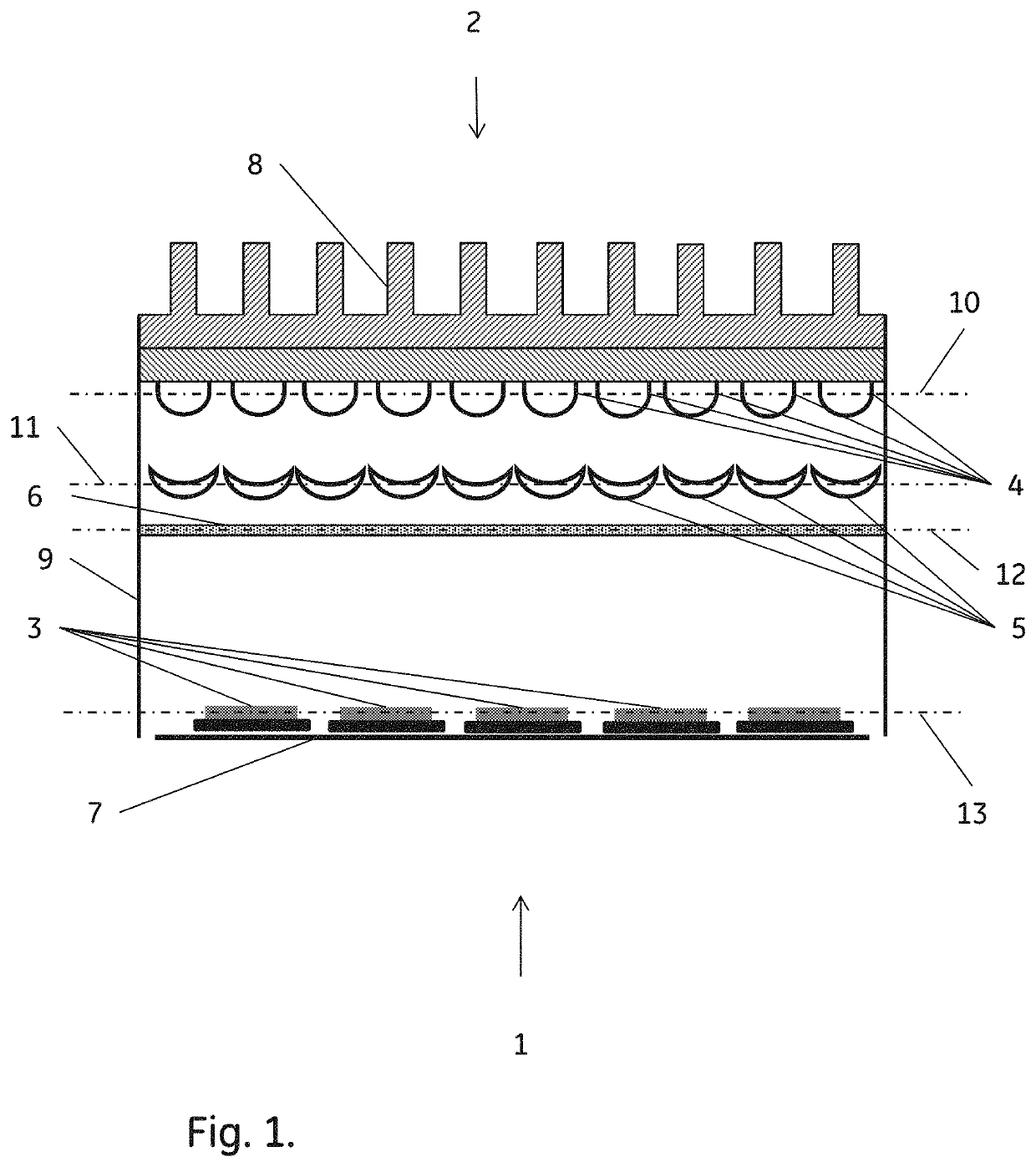

Image

  • Method for reduction of autofluorescence from biological samples
  • Method for reduction of autofluorescence from biological samples
  • Method for reduction of autofluorescence from biological samples

Examples


example 1.505

Example 1. 505 nm LED Light Treatment

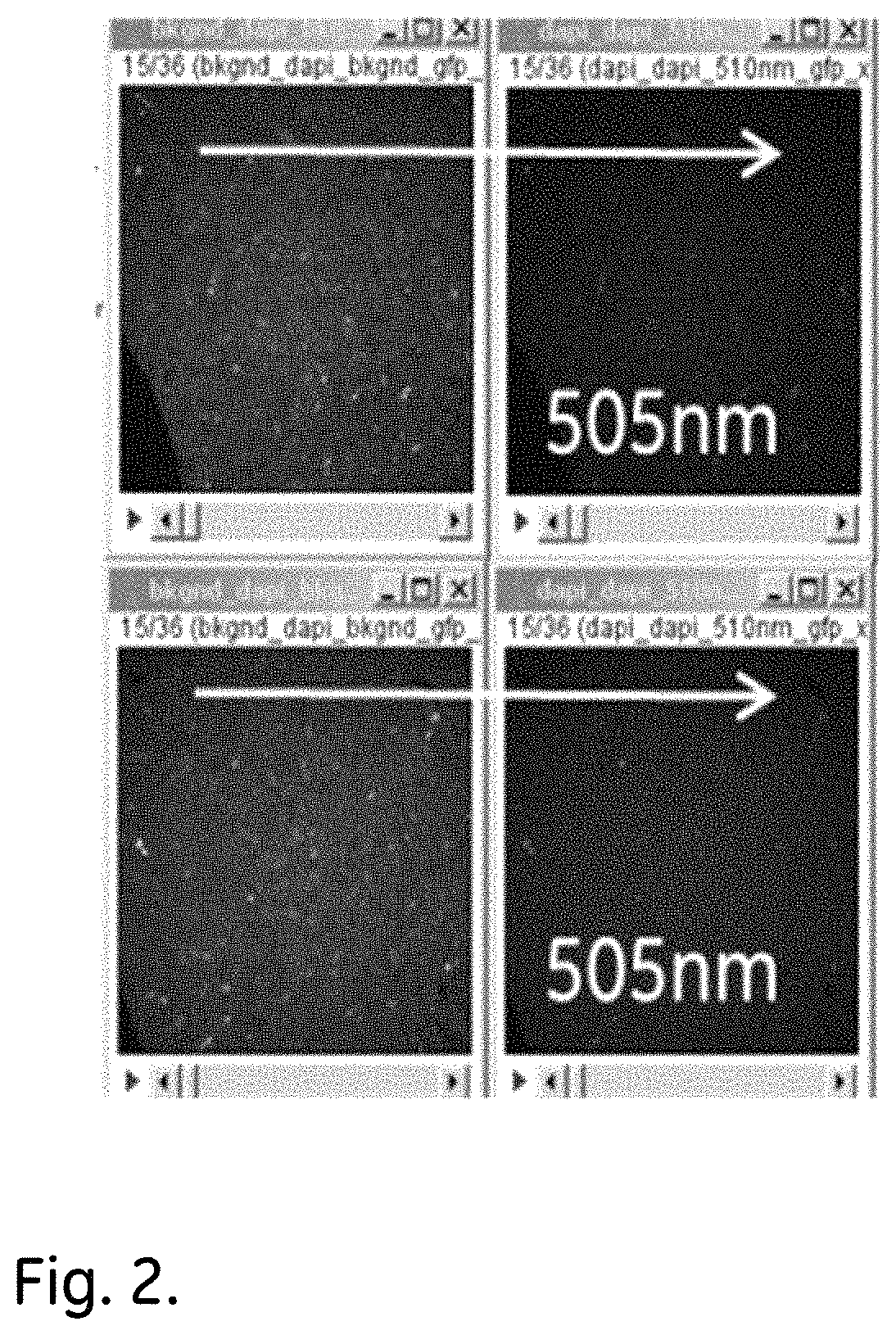
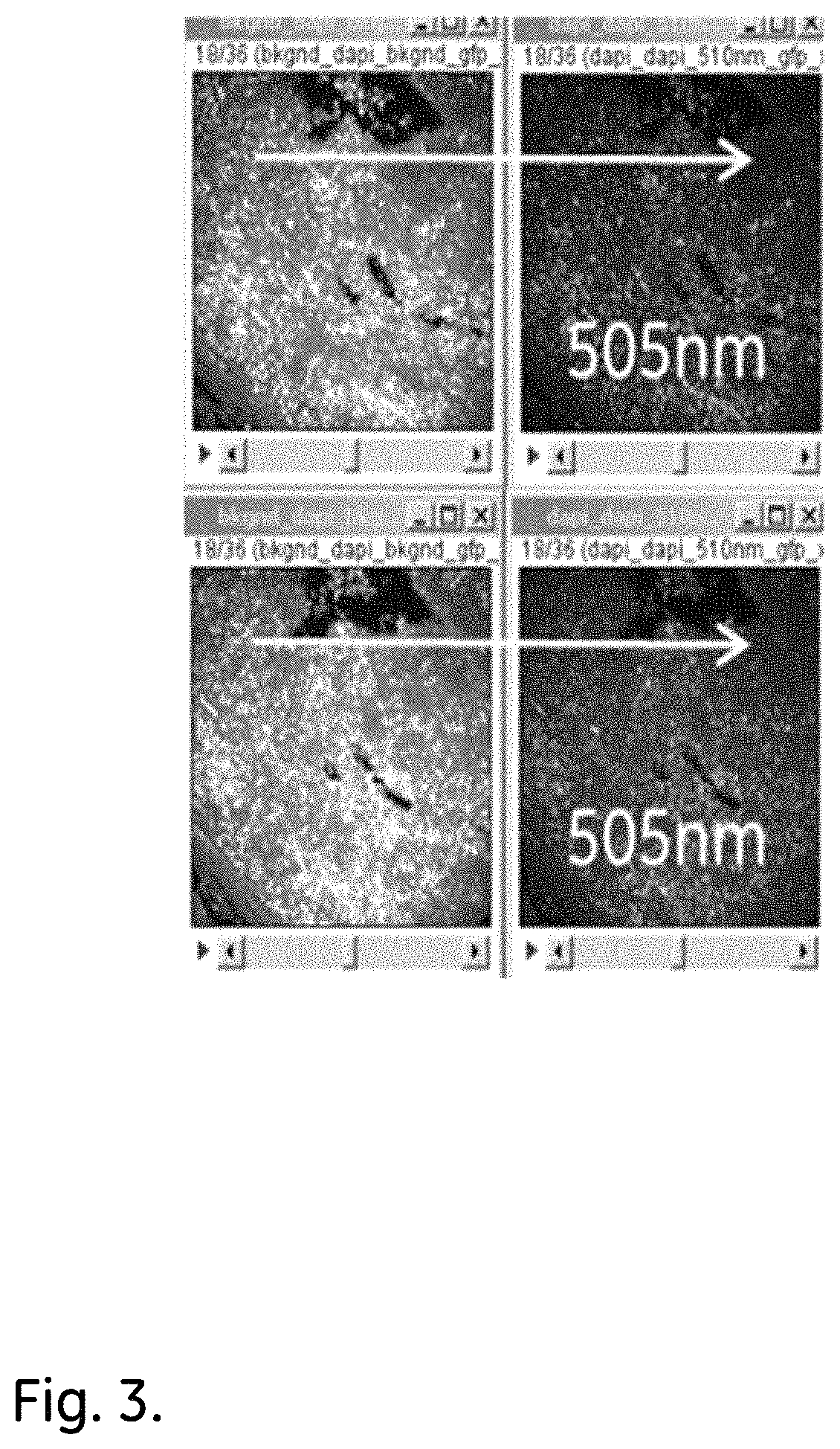
[0050]Deparaffinized sections of FFPE CHL (Classical Hodgkin Lymphoma) and T-cell lymphoma tissue samples were mounted on microscope slides and irradiated with 505 nm LED light, using a M505L3 LED, for 30 minutes.

[0051]The results, as shown in FIG. 2 (CHL) and 3 (T-cell) demonstrate that the light treatment is highly effective in AF reduction.

example 2

LED Wavelengths

[0052]Deparaffinized sections of an FFPE Folio T-cell Lymphoma tissue sample were mounted on microscope slides and irradiated for 30 minutes using 5 different LED wavelengths-385, 455, 505, 490 and 530 nm's. The LEDs used were M385LP1, M455L3, M505L3, M490L3 and M530L3.

[0053]The results, as shown in FIG. 4, demonstrate that a 30 min irradiation at 490, 505 or 530 nm gives effective AF reduction. LED with wavelengths above 455 nm show AF reduction whereas 385 nm LED exposure shows increase in AF.

example 3

LED Wavelengths

[0054]Deparaffinized sections of an FFPE Reactive lymph node tissue sample were mounted on microscope slides and irradiated for 30 minutes using 5 different LED wavelengths-385, 455, 505, 490 and 530 nm's.

[0055]The results, as shown in FIG. 5, demonstrate that 30 min irradiation at 490, 505 or 530 nm gives effective AF reduction, whereas 385 and 455 nm LED exposure shows increase in AF.


PUM

PropertyMeasurementUnit
widthaaaaaaaaaa
widthaaaaaaaaaa
wavelength intervalaaaaaaaaaa

Abstract

The invention discloses a method for reduction of autofluorescence in biological samples, comprising the steps of:a) providing a biological microscopy sample;b) irradiating the sample with visible light, wherein the visible light has a spectrum such that at least 50% of the light intensity emanates from a narrow wavelength interval within the visible range. The invention also discloses a method for autofluorescence reduction with triplet sensitizers irradiated with visible light.

Description

TECHNICAL FIELD OF THE INVENTION[0001]The invention relates generally to methods and systems for reducing inherent autofluorescence of biological materials in images of those biological materials and in particular to reduction of autofluorescence by photobleaching.BACKGROUND OF THE INVENTION[0002]Tissue auto-fluorescence (AF) is a fundamental problem in microscopy and surgical applications. It reduces the signal detection sensitivity, and in some cases may cause failure in the detection of fluorescent dye signals. Accurate detection of target-specific fluorescent dyes is critical for many microscopy applications, such as molecular pathology imaging, where quantitation of molecular pathways has significant implications such as predicting drug response, therapy planning, and population segmentation of cancer patients.[0003]In recent years the development of numerous fluorescent dyes has made optical fluorescent microscopy the method of choice for biomedical research. Numerous studies ...

Claims


Application Information

Patent Timeline
Patent Type & Authority Patents(United States)
IPC IPC(8): G01N21/64G01N33/574G01N33/483
CPCG01N21/6458G01N33/4833G01N33/57407G01N21/6428G01N2021/6439G01N2333/70503G01N2333/91215
Inventor SOOD, ANUPNATARAJAN, ARUNKUMARDYLOV, DMITRYKAANUMALLE, LAKSHMI SIREESHAMCDONOUGH, ELIZABETH MARY
Owner LEICA MICROSYSTEMS CMS GMBH
Learn More