

Method for reduction of autofluorescence from biological samples
a biological sample and autofluorescence technology, applied in the direction of fluorescence/phosphorescence, analysis by material excitation, instruments, etc., can solve the problems of reducing the detection sensitivity of signal detection, failure to detect fluorescent dye signals, and tissue autofluorescence (af) to achieve significant reduction of autofluorescence, rapid and easy to perform
- Summary
- Abstract
- Description
- Claims
- Application Information
AI Technical Summary
Benefits of technology
Problems solved by technology
Method used
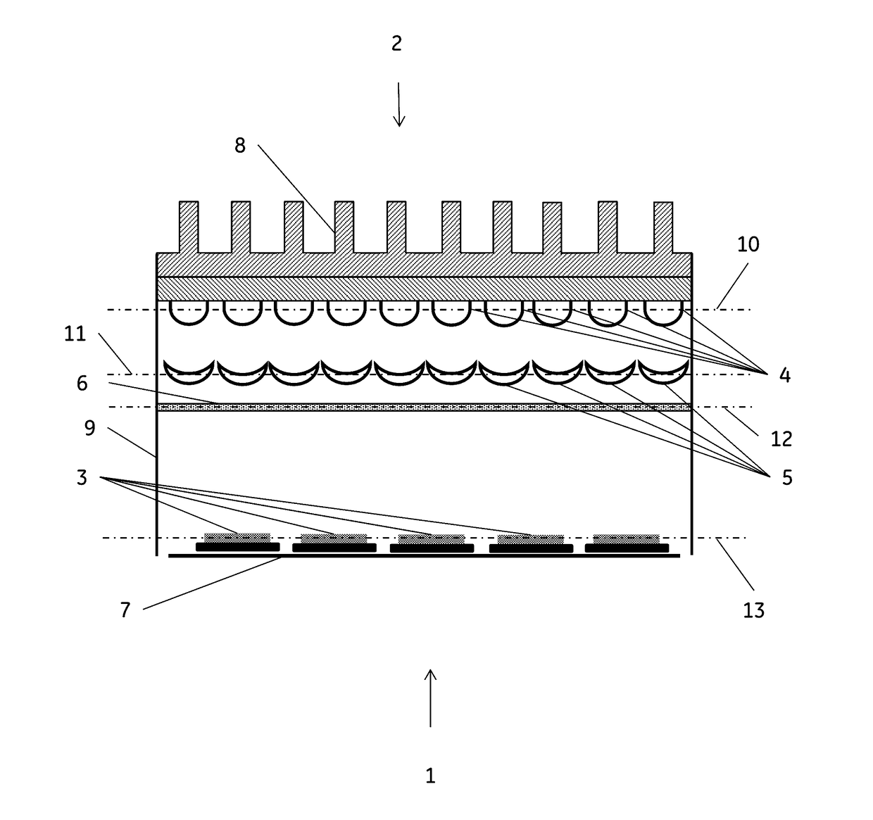
Image

Examples
example 1.505
Example 1. 505 nm LED Light Treatment
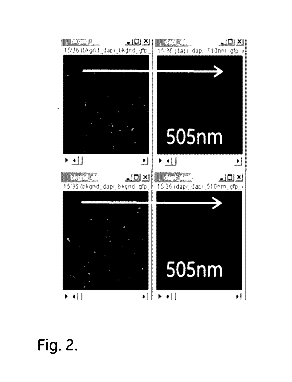
[0048]Deparaffinized sections of FFPE CHL (Classical Hodgkin Lymphoma) and T-cell lymphoma tissue samples were mounted on microscope slides and irradiated with 505 nm LED light, using a M505L3 LED, for 30 minutes.
[0049]The results, as shown in FIGS. 2 (CHL) and 3 (T-cell) demonstrate that the light treatment is highly effective in AF reduction.
example 2
LED Wavelengths
[0050]Deparaffinized sections of an FFPE Folio T-cell Lymphoma tissue sample were mounted on microscope slides and irradiated for 30 minutes using 5 different LED wavelengths-385, 455, 505, 490 and 530 nm's. The LEDs used were M385LP1, M455L3, M505L3, M490L3 and M530L3.
[0051]The results, as shown in FIG. 4, demonstrate that a 30 min irradiation at 490, 505 or 530 nm gives effective AF reduction. LED with wavelengths above 455 nm show AF reduction whereas 385 nm LED exposure shows increase in AF.
example 3
LED Wavelengths
[0052]Deparaffinized sections of an FFPE Reactive lymph node tissue sample were mounted on microscope slides and irradiated for 30 minutes using 5 different LED wavelengths-385, 455, 505, 490 and 530 nm's.
[0053]The results, as shown in FIG. 5, demonstrate that 30 min irradiation at 490, 505 or 530 nm gives effective AF reduction, whereas 385 and 455 nm LED exposure shows increase in AF.
PUM
| Property | Measurement | Unit |
|---|---|---|
| width | aaaaa | aaaaa |
| wavelength interval | aaaaa | aaaaa |
| wavelength interval | aaaaa | aaaaa |
Abstract
Description
Claims
Application Information