
Recombinant human serum transferrins containing peptides for inducing apoptosis in HIV-1 infected cells

a technology of transferrins and human serum, which is applied in the direction of transferrins, peptides, enzymology, etc., can solve the problem of unambiguous identification of donors

Inactive Publication Date: 2002-10-10
TOMYCZ NESTOR D
View PDF0 Cites 15 Cited by
  • Summary
  • Abstract
  • Description
  • Claims
  • Application Information

AI Technical Summary

Benefits of technology

[0021] The sequences flanking the RGD-containing peptides (bold, underlined) above are modified p17 / p24 HIV-1 protease cleavage sites (VSQNYVIVL). The natural p17 / p24 HIV-1 protease cleavage site is VSQNYPIVL, and HIV-1 protease cleaves at the YP junction. The modified p17 / p24 HIV-1 protease cleavage sites (VSQNYVIVL) used in the designed peptides have a proline to valine substitution in the P1' position of the cleavage site. This modification was used so that the peptides released following cleavage by HIV-1 protease have a valine (V) N-terminal terminal amino acid which is stabilizing according to the N-end rule; the mammalian N-end rule associates the intracellular half-life of a protein with its N-terminal amino acid. Cleavage of the modified p17 / p24 HIV-1 protease cleavage sites occurs at the YV junction, and the substitution of valine for proline has been shown to only have a minor effect on cleavage by HIV-1 protease. See Falnes et al. (1999) Biochem. J. 343:199-207. This modification substantially improves the stability of the released apoptosis-inducing peptides so that they are more likely to activate caspase-3 and the cell suicide pathway.
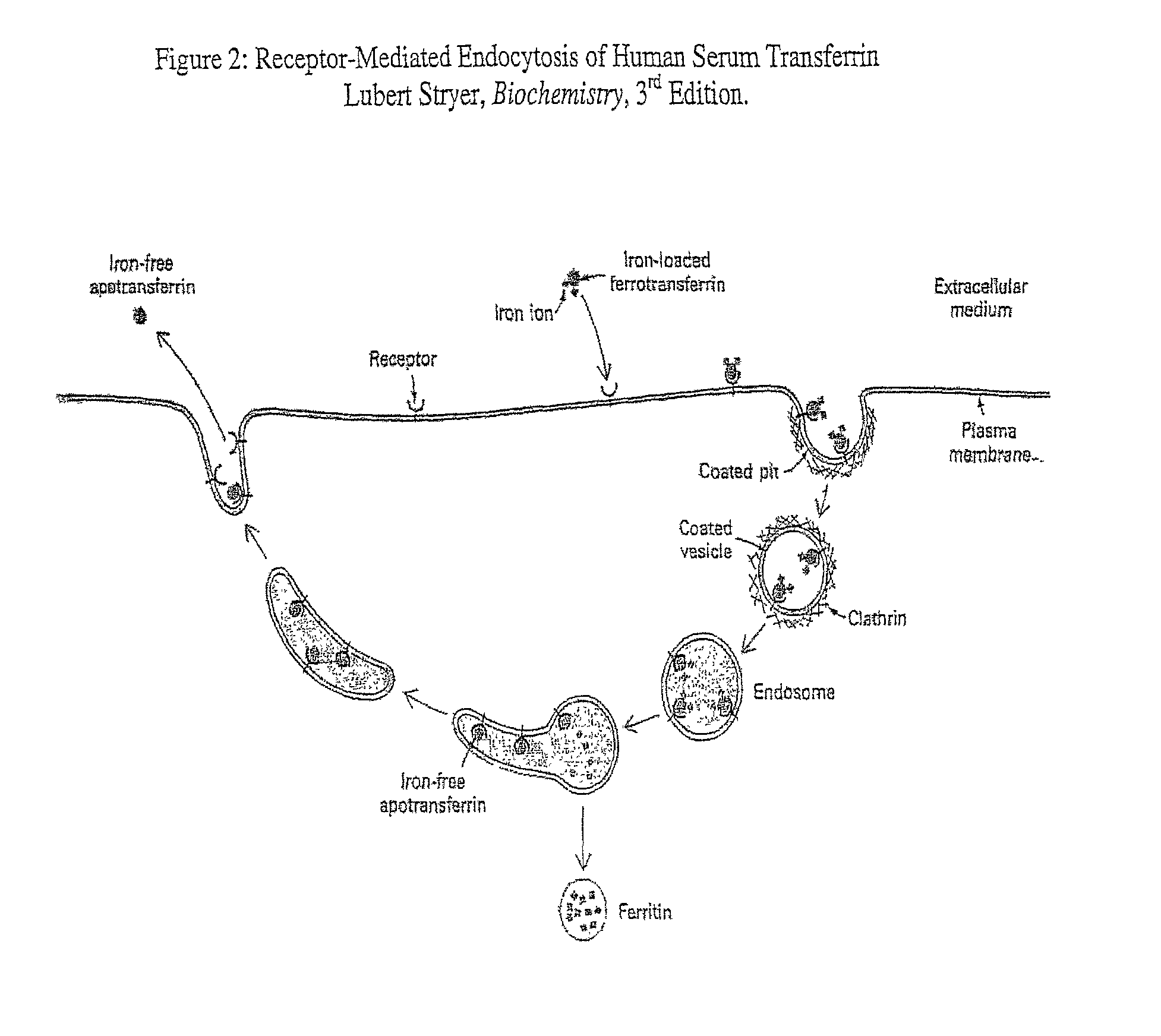
[0022] Those in the art will appreciate that human serum transferrin, a monomeric glycoprotein that binds tightly and reversibly to two ferric ions together with two bicarbonate co-ions, regulates the availability of free iron in body fluids, and also mediates the transport and cell uptake of iron by receptor-mediated endocytosis. See Huebers, H. et al. (1981) Proc. Natl. Acad. Sci. USA 78:1, 621-625; Jeffrey P. D. et al.(1998) Biochemistry 37:13978-13986; Ali, S. A et al. (1999) J. Biol. Chem. 274:34, 24066-24073. By this process, the transferrin molecule binds to the transferrin receptor on the membrane of cells and the complex is subsequently enveloped by the cell. Significantly, HIV replication has been shown to induce upregulation of HST receptor expression since HIV-infected cells exhibit a higher need for iron. See Levy, J. A. (1993) Microbiol. Rev. 57:183-289. Even more significantly, while conjugating peptides to human serum transferrin has been shown to induce an antibody immune response, hiding peptides within the loops of transferrin has not been associated with detectable antibody production. See Ali et al. (1999) FEBS Letters 459:230-232. Thus, human serum tranferrin provides an attractive pathway for the delivery of engineered peptides into HIV-infected cells as it takes advantage of a pre-evolved cell entry mechanism, exercises selectivity for HIV-infected cells which possess a greater number of transferrin receptors, and minimizes immunogenicity which often complicates the use of peptide drugs.
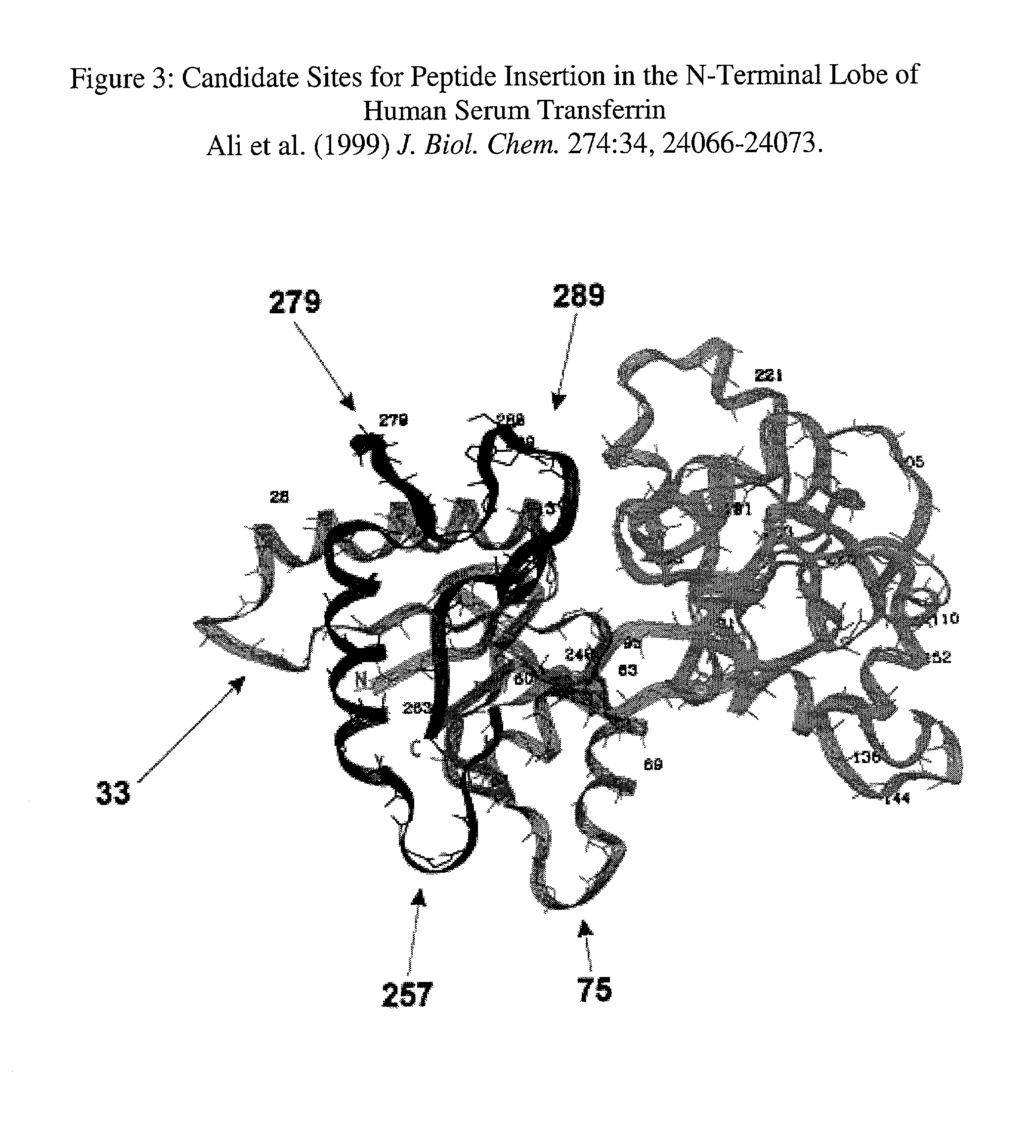
[0023] The efficacy of this mechanism is further enhanced by selecting insertion sites that are removed from the transferrin C-terminal lobe, known to be important for iron and bicarbonate binding. The C-terminal lobe of transferrin also possesses the primary sites for HST receptor recognition. See Zak et. al., J. Biol. Chem., 269:10, 7110-7114. Stability of the peptides once released into the cytoplasm is improved by including two modified p17 / p24 HIV-1 protease cleavage sites in substitution for the natural p17 / p24 cleavage sequence VSQNYPIVL. In this manner the released peptide conforms with the stabilizing N-end rule, and the peptide can be inserted into the N-terminal lobe surface exposed loops of transferrin, which are removed from the iron transport machinery. Surface exposed loops of the HST protein are the chosen targets of peptide insertion due to their flexibility and therefore resulting ability to tolerate insertions without significantly distorting the overall tertiary or three-dimensional spatial structure of the protein. See Finkelstein, A. V. (1997) Curr. Opin. Struct. Biol. 7:60-71. Furthermore, surface exposed loops are chosen so that peptide sequences can be exposed to HIV-1 protease recognition and cleavage.

Problems solved by technology

PCR is now also widely known as a forensic tool used to amplify the DNA of a single hair or sperm, leading to unambiguous identification of the donor.
Moreover, prior research has shown that insertion of peptides into the exposed loops of globular proteins is generally well tolerated without altering biological function, and such insertions do not significantly affect the stability and folding rates of the protein.

Method used


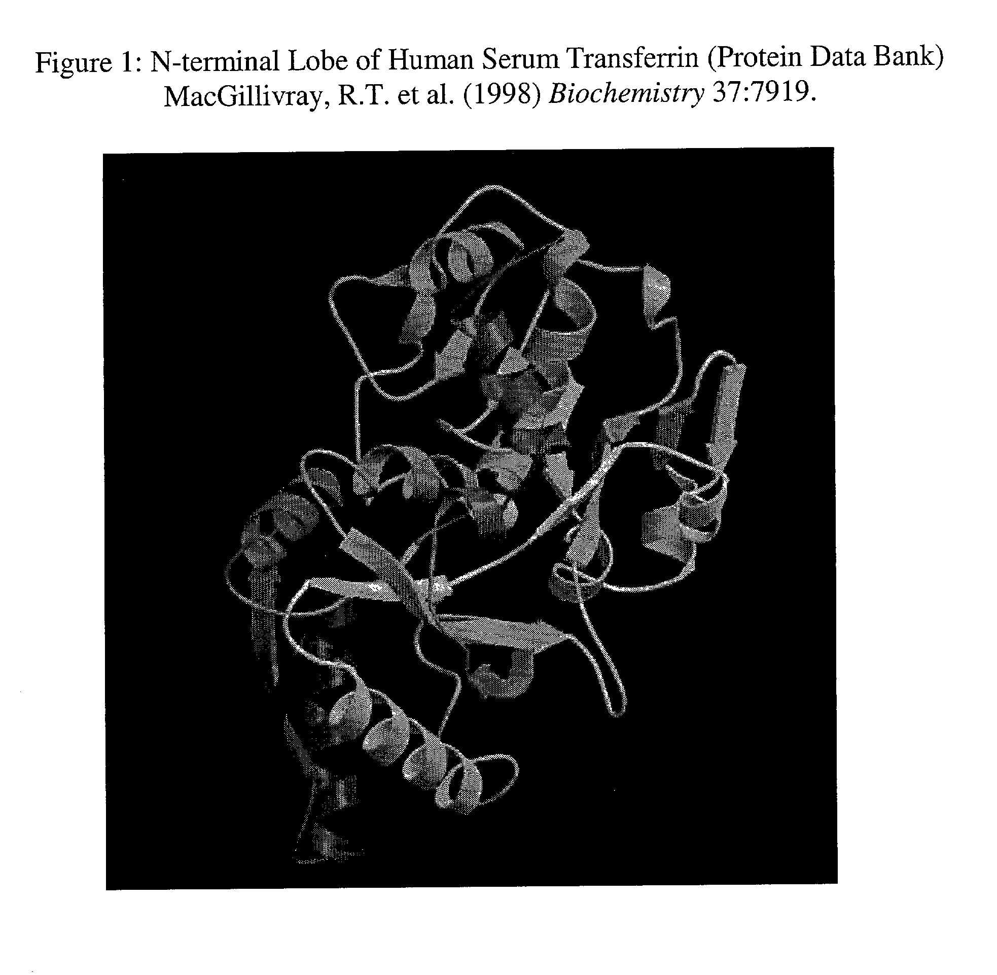
Image

  • Recombinant human serum transferrins containing peptides for inducing apoptosis in HIV-1 infected cells
  • Recombinant human serum transferrins containing peptides for inducing apoptosis in HIV-1 infected cells
  • Recombinant human serum transferrins containing peptides for inducing apoptosis in HIV-1 infected cells

Examples


example 1

[0047] VSQNYVIVLRGDVSQNYVIVL (Mut. 1),

Example 2

[0048] VSQNYVIVLRGDSVSQNYVIVL (Mut. 2),

example 3

[0049] VSQNYVIVLGRGDNPVSQNYVIVL (Mut. 3) and

example 4

[0050] VSQNYVIVLGRGDSPVSQNYVIVL (Mut. 4).

[0051] Each of the foregoing may be inserted at the N-terminal lobe sites hereinbefore described.

[0052] F. Subcloning of the HST Gene into the Baculovirus Protein Expression System:

[0053] The native HST gene would be subcloned into a baculovirus expression vector system (BEVS). The baculovirus expression vector system is now commonly used for expressing recombinant mammalian proteins. See O'Reilly, D. R., Miller, L. K., and Luckow, V. A. (1994) Baculovirus Expression Vectors: A Laboratory Manual, Oxford Univ. Press, Oxford. The baculovirus expression vector system uses insect cells to produce protein, and it is the first non-mammalian expression system that has been used successfully to generate functional full-length HST proteins. See Ali et al. (1996), Biochem. J. 319:101-105. The baculovirus expression method of Ali et al. (1996) produced a high-yield of functionally active HST (>20 mg / L), and this general method will be utilized for expre...


PUM

PropertyMeasurementUnit
Concentrationaaaaaaaaaa
Protein structureaaaaaaaaaa

Abstract

A mammalian protein, like the human serum transferrin (HST), is modified with an inserted peptide sequence flanked at both ends by cleavage sites. The peptide insert contains a motif known to induce apoptosis in cells and the cleavage sites are specific for the viral protease of HIV-1. The delivery of such recombinant transferrin into an HIV-1 infected cell results in the release of the peptide which then induces apoptosis. The peptide is inserted into surface exposed loops of the N-terminal lobe of the HST containing the RGD motif flanked by two modified p17 / p24 HIV-1 protease cleavage sites. When delivered to the infected cell the cleavage of the loop inserted sequences by the HIV-1 protease results in the release of the central RGD-containing peptide sequences. Peptides containing the RGD motif (arginine, glycine, aspartic acid) have been shown to induce cell apoptosis even in small concentrations.

Description

[0001] 1. Field of the Invention[0002] The present invention relates to biological structures designed to induce apoptosis in HIV-1 infected cells, and more specifically to recombinant human serum transferrins designed to introduce apoptosis-inducing peptides into HIV-1 infected cells.[0003] 2. Description of the Prior Art[0004] The preservation of equilibrium in a multicellular organism, the process of homeostasis, requires a delicate balance between cell proliferation and cell death. In a well developed, adult organism the maintenance and renewal of many specialized cell lines is a matter of continuing importance. Therefore, cell proliferation and in particular its tight checks and balances necessary for optimal adult health has been a process of keen interest to those involved in biological research. Accordingly, the study of infectious diseases and more recently the focused inspection of mechanisms associated with cancer have followed these adult self-preservation paths, expandi...

Claims


Application Information

Patent Timeline
IPC IPC(8): A61K38/00A61K38/40C07K14/16C07K14/79C07K14/82C12N15/62C12P21/04
CPCA61K38/40C07K14/005C07K14/79C07K14/82C12N2740/16222C07K2319/035C07K2319/50C07K2319/75C12N15/62C07K2319/00
Inventor TOMYCZ, NESTOR D.
Owner TOMYCZ NESTOR D
Learn More