

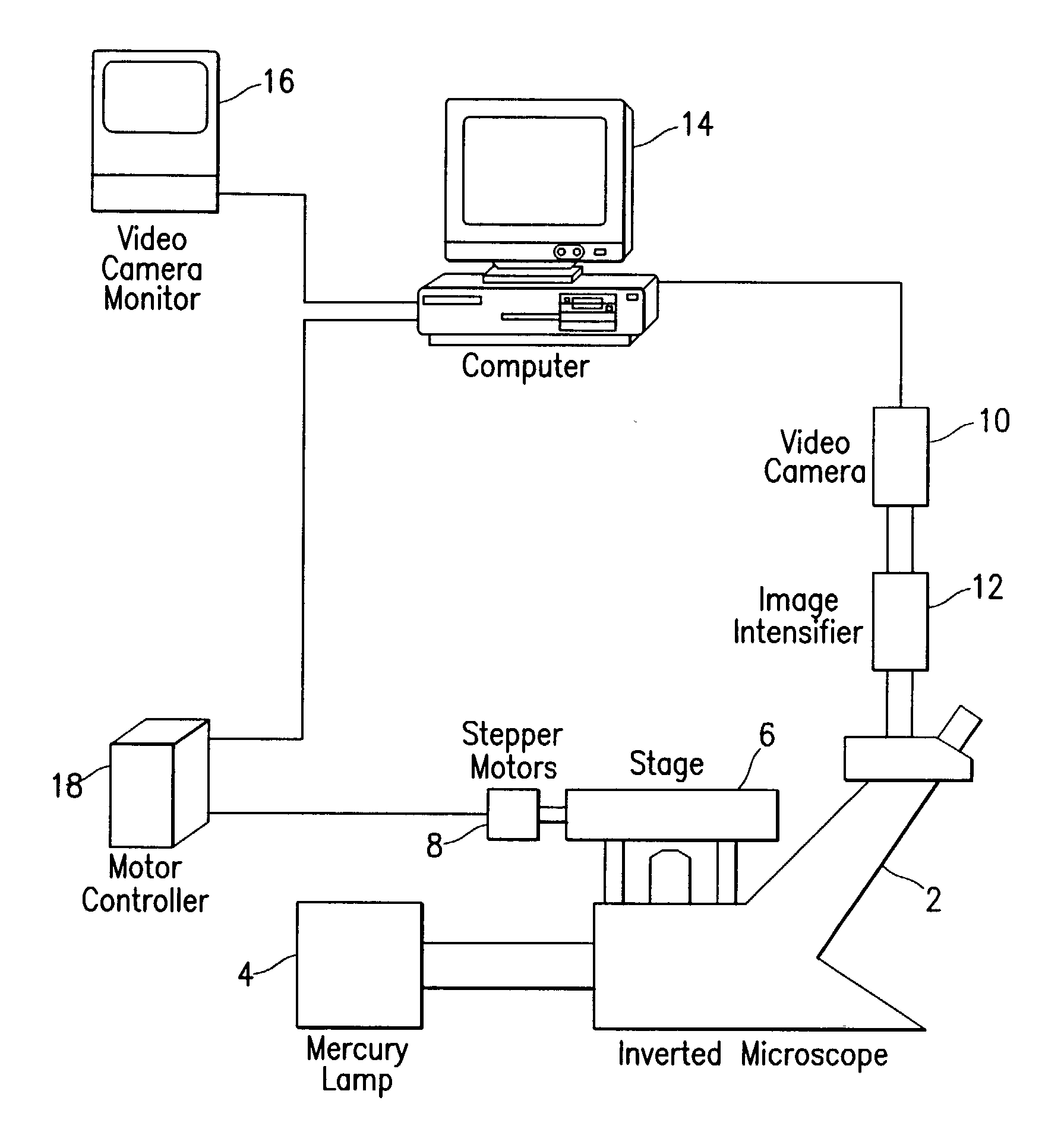
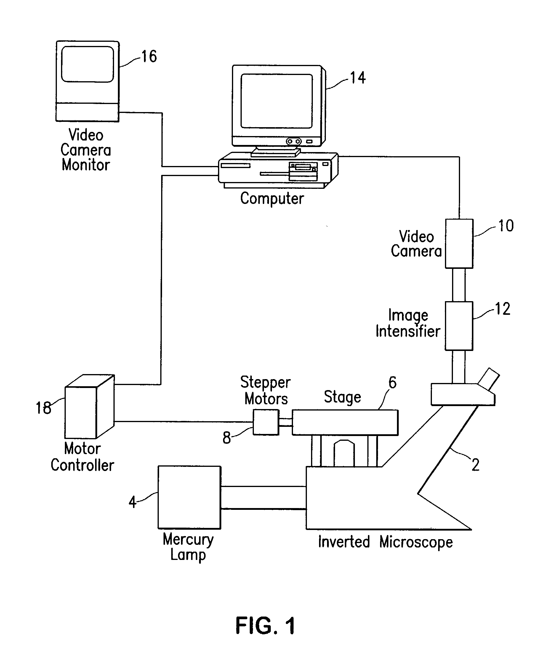
Fluorescence digital imaging microscopy system
a digital imaging and microscopy technology, applied in the field of tissue culture container relative cell number quantification system, can solve the problems of limited flexibility of existing fluorescence readers, radioactivity risk, and inability to provide accurate and reliable results,
- Summary
- Abstract
- Description
- Claims
- Application Information
AI Technical Summary
Benefits of technology
Problems solved by technology
Method used
Image

Examples
example 1
[0061] Initial tests for the linearity and sensitivity of the preferred embodiment of the fluorescence digital imaging microscopy system were performed on cells which were treated with dye separate from the 96-well plate, to minimize any effects from loading dye in situ. BA-1 hybridoma cells (which grow as a single cell suspension) were incubated with BCECF-AM in a test tube and rinsed with RPMI-1640+10% FCS prior to loading in a 96-well plate. FIG. 9 shows the serial dilution curve. Fluorescence at 4.times. magnification was very linear over a range from 340 cells / well through 350,000 cells / well (3.4.times.10.sup.3 cells / ml to 3.5.times.10.sup.6 cells / ml), with a coefficient of linearity equal to 0.995. The system provided excellent linearity covering approximately 3 logs of fluorescence and 3 logs of cell density.
example 2
[0062] For initial testing of in situ linearity, Hoechst 33342 dye was used because it produces minimal background fluorescence. FIG. 10 shows a typical dilution curve for SMS-KCNR, a neuroblastoma cell line, in RPMI-1640+10% FCS treated in situ with Hoechst 33342 (10 .mu.g / ml for 30 min. at 37.degree. C.) in a 96-well plate and scanned at 4x magnification without an image intensifier. Good linearity (r=0.997) was seen from 1.times.10.sup.3 cells / well to 5.4.times.10.sup.5 cells / well (1.0.times.10.sup.4 cells / ml to 5.4.times.10.sup.6 cells / ml). Results were equally good at magnifications of 10.times. and 4.times. with image intensification, producing coefficients of linearity of 0.997 and 0.994 respectively.
example 3
Eosin Y Treatment of Samples Containing Large Numbers of Non-viable Cells
[0063] Serial dilution of viable cells. For studies of the correlation of relative fluorescence and viable neuroblastoma cell number, cells were loaded in Falcon 96-well tissue culture plates (Becton-Dickinson, Lincoln Park, N.J.) with an Electrapette multi-channel pipettor (Matrix Technologies, Lowell, Mass.) set for serial twofold dilution mixing 125 .mu.l volume in 3 cycles. One million cells per well to two cells per well were plated in 8 replicate wells per condition. Final well volume was 125 .mu.l. Neuroblastoma cells were allowed to settle and attach for 2-8 hours. Prior to scanning, the tissue culture plates were gently loaded by multi-channel pipettor with 50 .mu.l per well of intermediate FDA stock and incubated at 37.degree. C. for 30 minutes. After incubation, plates were loaded with 30 .mu.l per well of 0.5% eosin Y solution, yielding final eosin Y concentration of 0.083%; controls received a medi...
PUM
| Property | Measurement | Unit |
|---|---|---|
| concentration | aaaaa | aaaaa |
| concentration | aaaaa | aaaaa |
| volume | aaaaa | aaaaa |
Abstract
Description
Claims
Application Information