Rapid screening procedure for inflammation mediators

a screening procedure and inflammation mediator technology, applied in the field of rapid screening procedure of inflammation mediators, can solve the problems of toxic effects, potential adverse effects of cell free oxyhb substitutes, patients with activated immune systems,

Inactive Publication Date: 2003-06-26
CASSIDY RICHARD A +3
View PDF4 Cites 1 Cited by
  • Summary
  • Abstract
  • Description
  • Claims
  • Application Information

AI Technical Summary

Benefits of technology

0068] Total nitrate and nitrite levels in filtrates of PMNs were determined by reducing the nitrate to nitrite by nitrate reductase and reacting the resulting nitrite with 2,3-diaminonaphthalene to form a fluorescent product. PMNs (500,000/300 ul HBSS) were treated with oxyMb (1-1000 82 g/mL) and SOD (30 U) for 10 min followed by PMA (200 nM) for 1 h at 30.degree. C. Filtrates were collected by centrifugation at 16,000 g for 15 min through Ultrafree-MC 10,000 NMWL filters (Millipore, Bedford, Mass.). Total nitrite levels were determined with a 96-well fluorescence analyzer (IDEXX, Westbrook, Me.). PMNs exposed to the same treatment were analyzed by flow cytometry for DCFH oxidation.
0069] OxyHb bound to and internalized into PMNs was determined using fluorescein-conjugated oxyHb (Molecular Probes, Eugene, Oreg.). Each molecule of chromatographically purified oxyHb-A.sub.0 (98% ferrous) had an average of 4.5 molecules of fluorescein-5-EX succinimidyl ester conjugated to lysine (oxyHb-FLEX). OxyHb-FLEX (1-250 .mu.g/mL) was incubated with PMNs with and without albumin (20 mg/mL) for 10 min in a shaking water bath at 37.degree. C. followed by a 30 min incubation with or without LPS (10 ng/mL) and TNF.alpha. (1 ng/mL) or PMA (200 nM). Flow cytometric measurements were performed as described above. To visualize intracellular levels of oxyHb-FLEX, whole cell and Z-plane images of PMNs were collected using an Axiovert 135 inverted confocal microscope (Zeiss, Thornwood, N.Y., and Atto Instruments, Rockville, Md.).
0070] DCFH-loaded PMNs were incubated for 10 min at 37.degree. C. with various amounts of oxyMb (1-1000 .mu.g/mL) with or without L-NMMA (5 mM), N-(1-iminoethyl)-L-ornithine (L-NIO) (5 mM), ascorbate (25 .mu.M) and glutathione (25 .mu.M), human albumin (20 mg/mL), or human ery...

Problems solved by technology

This decreases extracellular O.sub.2..sup.-, allowing NO. concentrations to rise, enter neighboring cells, and produce toxic effects.
In summary, disease processes that release heme proteins may result in the binding of heme protein to activated PMN...

Method used


Image

  • Rapid screening procedure for inflammation mediators
  • Rapid screening procedure for inflammation mediators
  • Rapid screening procedure for inflammation mediators

Examples


example 1

PMNs and Lymphocyte Isolation

[0063] All reagents, unless otherwise specified, were obtained from Sigma Chemical Co. (St. Louis, Mo). Human PMNs and lymphocytes were obtained from EDTA-preserved venous blood of non-smoking adult males by layering blood over Polymorphprep, Nycomed Pharma As (Oslo, Norway). Blood was centrifuged at 550 g for 30 min at 22.degree. C., and the PMN and lymphocyte layers were collected and washed twice in Hanks' balanced salt solution (HBSS) Gibco (Grand Island, N.Y.). All PMNs were incubated with physiological levels (10.sup.-4 M) of arginine unless otherwise stated. One or two hypotonic lyses were performed to lower the red blood cells to .ltoreq.1% of PMNs or of lymphocytes. RBC ghosts lying on top of the PMNs were removed after the first hypotonic lyse by pipette extraction. The purity of PMNs and lymphocytes was greater than 88% and 95%, respectively, and their viability as determined by trypan blue-uptake was greater than 90%. All experiments were con...

example 2

Effect of NO. or H.sub.2O.sub.2 Concentration or PMN Density on DCFH Oxidation

[0064] PMNs were incubated with 2,7-dichlorodihydrofluorescein diacetate (DCFH-DA; (2 .mu.M; from Kodak, Rochester, N.Y.) at 37.degree. C. for 15 minutes. DCFH-DA permeated the cells freely and was trapped after enzymatic hydrolysis of the diacetate to DCFH. Oxidation of DCFH resulted in the fluorescent DCF. To assess the use of DCFH-DA in measuring intracellular levels of NO and / or H.sub.2O.sub.2, a NO. donor, diethylamine nitric oxide (DEANO; Molecular Probes, Eugene, Oreg.) or hydrogen peroxide (H.sub.2O.sub.2), at concentrations of (10.sup.-11 to 5.times.10.sup.-4 M) were incubated with DCFH-loaded PMNs at 37.degree. C. for 30 min. Concentrations of DEANO-derived NO. were generated by rapidly performing an initial 10-fold serial dilution of a concentrated (10.sup.-2 M), alkali (1 mM NaOH) stock into HBSS buffer followed by a 10 fold dilution into the PMN preparations. Intracellular levels of propidium ...

example 3

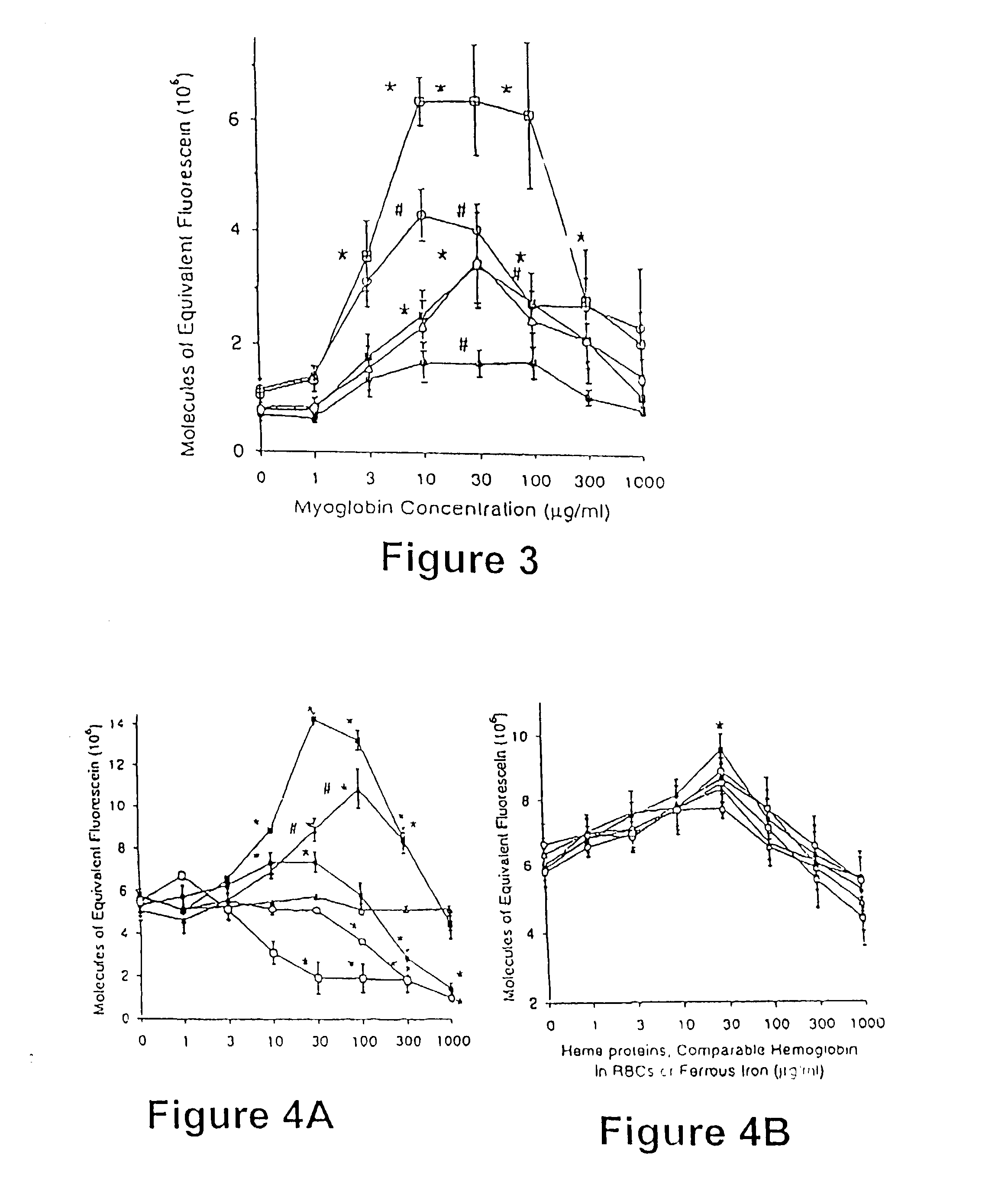
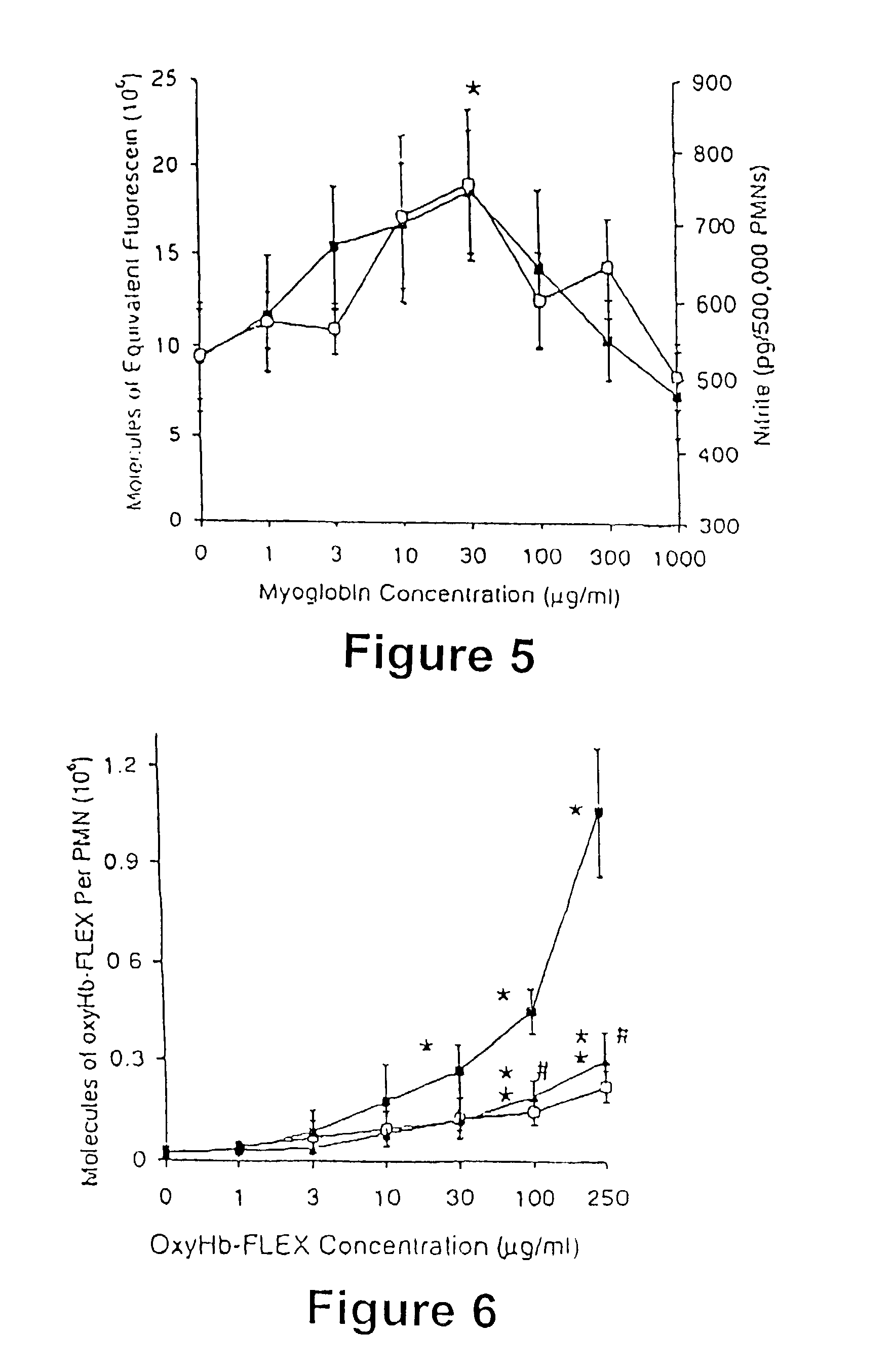
Heme Protein Effect on DCFH Oxidation in Unstimulated, Lipopolysaccharide (LPS) and Tumor Necrosis Factor .alpha. (TNF.alpha.)-, and PMA-stimulated PMNs

[0066] DCFH-loaded PMNs were incubated with and without N-methyl-L-arginine (L-NMMA; 5 mM; Calbiochem, San Diego, Calif.) and heme proteins (0.3-1000 .mu.g / mL) at 37 .degree. C. for 10 minutes. The L-NMMA-treated PMNs were subsequently incubated for an additional 30 min in the absence or presence of LPS (10 ng / mL), LPS (10 ng / mL) and TNF.alpha. (1 ng / mL), or PMA (200 nM). In unstimulated and LPS and TNF.alpha. -stimulated PMNs, the treatments were oxyMb, or oxyMb+L-NMMA (5 mM). In PMA-stimulated PMNs with arginine, the treatments were oxyMb, oxyHb, bilirubin, ferrous chloride or either isolated RBCs or sonicated RBCs containing comparable amounts of oxyHb. RBC cell membranes were ruptured using a micro-ultrasonic cell disrupter, Kontes, Janke and Kukel Gmbh and CO. (Stauten, Germany). In PMA-stimulated PMNs without arginine, the trea...


PUM


Abstract

The invention is compositions and methods for determining the presence of one or more immune response mediators, comprising obtaining a solution containing polymorphonuclear neutrophils, exposing the neutrophils to a chromophore, allowing the chromophore to oxidize to form a luminescent compound, and measuring the level of visible or ultraviolet radiation.

Description

(B) CROSS-REFERENCE TO RELATED APPLICATIONS[0001] This application is a continuation-in-part of PCT / US01 / 07434, filed Mar. 9, 2001, which claims priority from U.S. Serial No. 60 / 272,048, filed Mar. 1, 2001 and U.S. Serial No. 60 / 188,001, filed Mar. 9, 2000.(C) STATEMENT REGARDING FEDERALLY SPONSORED RESEARCH[0002] Not applicable.(D) BACKGROUND OF THE INVENTION[0003] (D1) Field of the Invention[0004] The present invention is directed to a screening procedure and compositions for rapidly evaluating drugs that mediate inflammation.[0005] (D2) Description of Related Art[0006] Severe tissue injury, rhabdomyolysis, and myocardial infarction result in the release of myoglobin (Mb) from muscle into the interstitium and the vasculature. Hemoglobin (Hb) is released from erythrocytes during hemolytic disorders, burns, and storage of blood for transfusion. In humans, elevated levels of Mb or Hb have been reported to be associated with acute renal failure, infections, and recurrent cancers in po...

Claims


Application Information

Patent Timeline
IPC IPC(8): G01N33/50G01N33/68
CPCG01N33/6863G01N33/5047
Inventor CASSIDY, RICHARD A.BURLESON, DAVID G.SALIN, MARVIN L.DELGADO, ANGEL V.
Owner CASSIDY RICHARD A
Why Eureka
Try Eureka