
Optimized high-speed magnetic resonance imaging method and system using hyperpolarized noble gases

a high-speed magnetic resonance imaging and noble gas technology, applied in the direction of magnetic variable regulation, process and machine control, instruments, etc., can solve the problems of high temporal resolution, no extension, and no means to visualize gas-flow dynamics with high spatial resolution, and no extension, and no extension, can solve the problem of high spatial resolution, high temporal resolution, and low resolution of high-speed conventional gradient-echo pulse sequences. the effect of high spatial resolution

Active Publication Date: 2004-12-23
UNIV OF VIRGINIA ALUMNI PATENTS FOUND +1
View PDF4 Cites 28 Cited by
  • Summary
  • Abstract
  • Description
  • Claims
  • Application Information

AI Technical Summary

Benefits of technology

[0018] The present invention comprises the method, related system, and computer useable medium (readable media) for using hyperpolarized noble gases together with an appropriately designed and optimized MRI pulse sequence to yield a technique that: (1) generates images with high spatial resolution, high temporal resolution, and negligible image artifacts, and (2) permits, in appropriate applications, the high temporal resolution to be easily traded for increased spatial coverage.
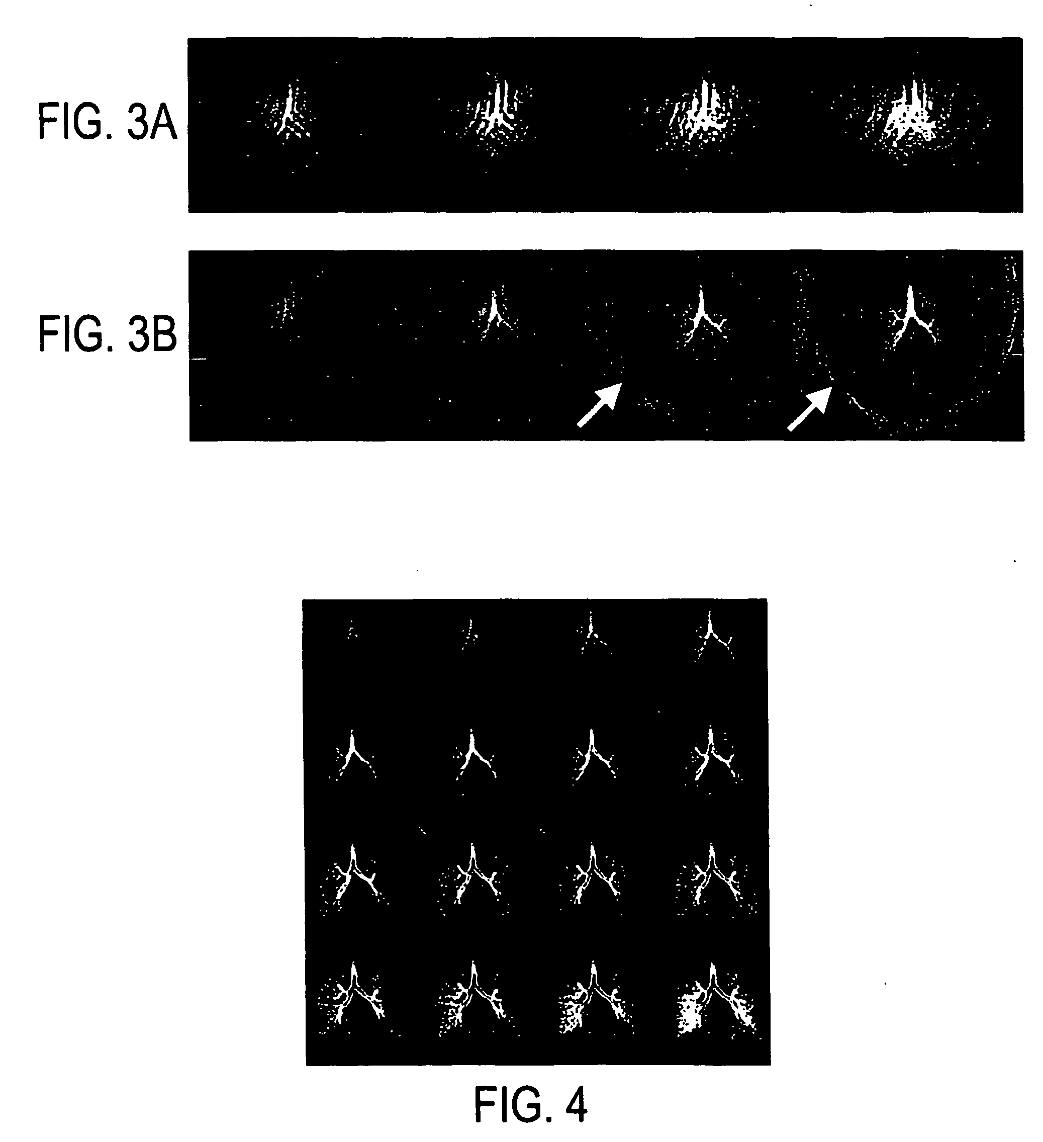
[0020] An important advantage and application of the present invention is the visualization of gas-flow dynamics. Since the present invention method permits gas-flow patterns to be determined without the introduction of secondary tracer materials and without the need to have direct visual contact with the flowing gas, it presents the opportunity to perform, in a variety of fluid systems, measurements of gas-flow dynamics that were heretofore not possible. One specific example is the possibility to dynamically image, in vivo, the ventilatory function of the human or animal lung, thus providing information on the physiology of normal lung finction or the pathophysiology of lung disease that cannot be obtained by any other means.

Problems solved by technology

However, all of these previous attempts have notable limitations, and none, nor any of their extensions, provide the means to visualize gas-flow dynamics with high spatial resolution, high temporal resolution, and negligible image artifacts.
Rapid conventional gradient-echo pulse sequences lack the desired temporal resolution.
Yet conventional gradient-echo imaging is undesirable in this respect as an RF pulse is required for each line of spatial-frequency-space (k-space) data.
Although EPI provides efficient use of the magnetization and high temporal resolution, it is prone to signal loss and / or geometric distortion secondary to the effects of magnetic field inhomogeneities during the relatively long data-sampling period.
In addition, the gradient-intensive nature of EPI makes it susceptible to diffusion-induced signal losses when gases are imaged.
Additionally, slice orientations other than axial demonstrate substantial bulk magnetic susceptibility-induced image distortions due to the structure of the lung, thus reducing the utility of the technique.
Furthermore, the EPI pulse-sequence structure requires an echo time that is on the order of the T2*, resulting in marked signal loss around vessels due to susceptibility effects (See Saam, Yablonskiy, et al.).
However, an attempt at dynamic hyperpolarized-gas imaging using interleaved-EPI resulted in substantial motion artifacts during periods of rapid gas flow (See Ruppert, Brookeman et al.).
However, the total acquisition time is generally limited, for example by the spin-lattice (T1) relaxation time of the hyperpolarized gas, or, for applications such as human lung imaging, by the subject's ability to suspend respiration.
Also, while traditional commercial methods provide linear gradients in the x, y, or z directions it is also possible not to utilize all three of these linear gradients.
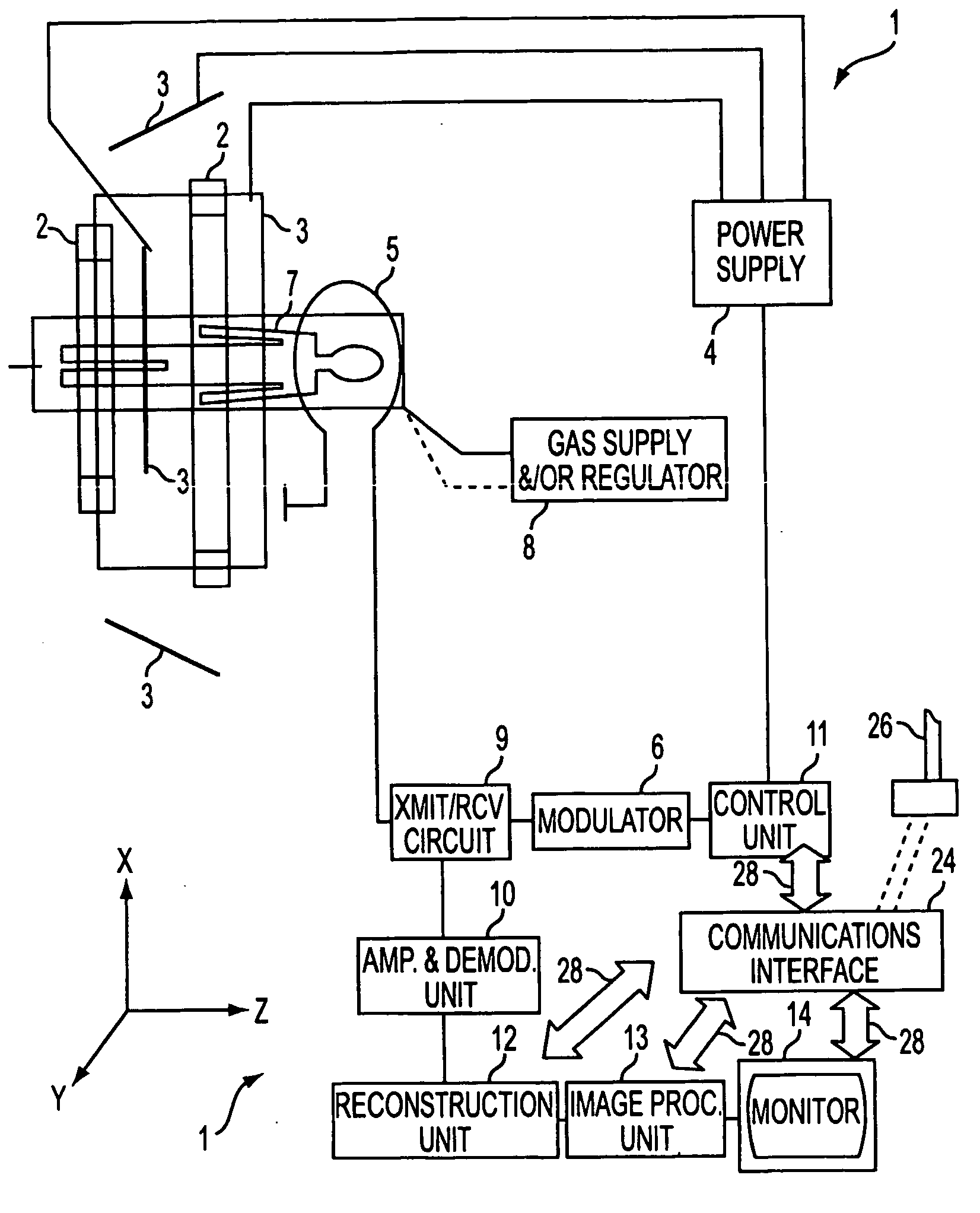
In particular, the control unit commanded by its loaded software causes the generation of MR signals by controlling the application of MR pulse sequences, which comprise RF-pulses, time delays and temporary magnetic-field gradient pulses.

Method used


Image

  • Optimized high-speed magnetic resonance imaging method and system using hyperpolarized noble gases
  • Optimized high-speed magnetic resonance imaging method and system using hyperpolarized noble gases
  • Optimized high-speed magnetic resonance imaging method and system using hyperpolarized noble gases

Examples


example no.1

EXAMPLE NO. 1

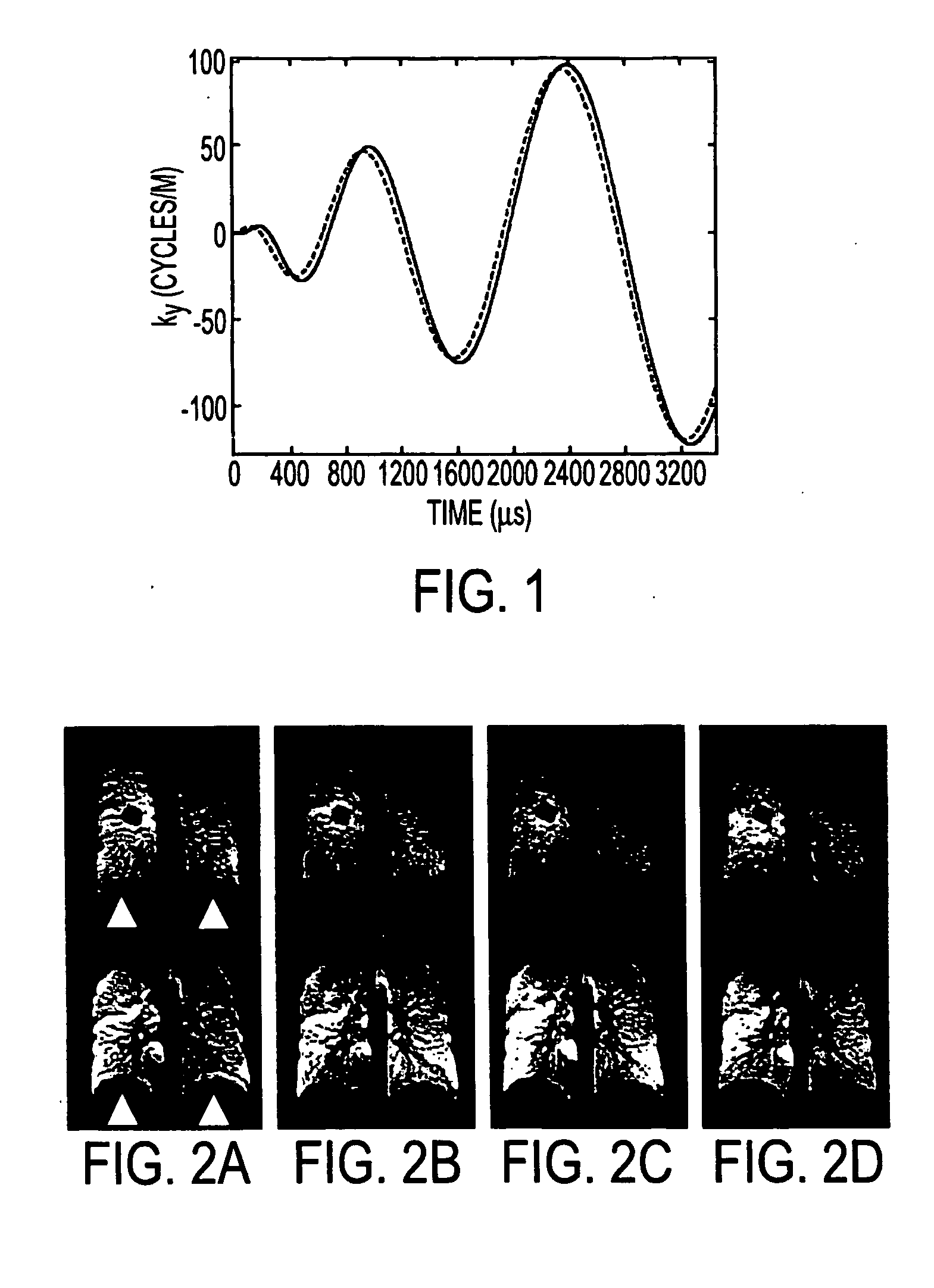
[0096] A specific pulse-sequence implementation that satisfies the six general requirements listed above is useful to illustrate the nature of the invention. For this purpose, we will discuss the case of an interleaved-spiral-type pulse sequence (See Meyer C. H., Hu B. S., Nishimura D. G., Macovski A., "Fast Spiral Coronary Artery Imaging", Magn. Reson. Med., 1992, 28:202-213 [herein after "Meyer, Hu et al."]). This k-space trajectory was chosen as a starting point because, by nature, an interleaved spiral satisfies requirements (1), (2), (3) and (4). The interleaved-spiral trajectories were designed specifically for .sup.3He imaging and were optimized specifically to meet requirement (6). Further, we demonstrated that these optimized trajectories meet requirement (5) for .sup.3He imaging. In addition, we implemented a specific temporal ordering for the acquisition of the spiral trajectories that further decreases the level of motion artifacts.

[0097] We have used the re...

example no.2

EXAMPLE NO. 2

[0144] Dynamic Imaging in Patients with Lung Disease

[0145] We tested the dynamic interleaved-spiral imaging technique in a variety of lung pathologies including asthma, cystic fibrosis, chronic obstructive pulmonary disease, emphysema secondary to .alpha.-1 antitrypsin deficiency, and post lung transplant. The patients were instructed to inhale the gas over the first half, and exhale over the second half, of the 15-second acquisition period. Imaging was performed in the coronal orientation.

[0146] Referring to FIG. 8A, in a patient with severe asthma there were several regions that demonstrated abnormal ventilation patterns. Since this subject's lungs were hyperinflated, as typically seen in this disease, there was limited diaphragmatic excursion during inspiration. The images demonstrated a heterogeneous filling pattern, with portions of the middle lung zones filling prior to the lower lung. It was also evident that some regions of the lung filled at different rates and...

example no.3

EXAMPLE NO. 3

[0154] Introduction

[0155] Hyperpolarized .sup.3He spin-density imaging of the lung shows promise for characterizing diseases such as asthma and emphysema. As the size, location and shape of ventilation defects resulting from these diseases are highly variable, volumetric imaging with isotropic spatial resolution is desirable for their accurate quantification.

[0156] Hyperpolarized .sup.3He imaging has most commonly used gradient-echo (GRE) pulse sequences. However, volumetric GRE imaging with isotropic spatial resolution of a few millimeters requires an acquisition time of .about.20 seconds, which is longer than the breath-hold duration that can be tolerated by patients with compromised respiratory function.

[0157] A goal of this investigation was to develop interleaved-spiral pulse sequences using optimized slew-limited spirals to obtain images covering the whole lung with 4-mm isotropic spatial resolution in under 10 seconds. We compared two strategies: a rapid 2D inter...


PUM


Abstract

A system and method for using hyperpolarized noble gases together with an appropriately designed and optimized magnetic resonance imaging pulse sequence to rapidly acquire static or dynamic magnetic resonance images. The strong magnetic resonance signal from hyperpolarized gases, combined with the present magnetic resonance imaging technique, presents the opportunity for the imaging of gases with both high spatial and high temporal resolution. One potential application for such a method is the direct, dynamic visualization of gas flow, which would be extremely useful for characterizing a variety of fluid systems. In the medical field, one such system of substantial importance is the lung. The system and method provides for visualizing regional ventilatory patterns throughout the respiratory cycle with high temporal and high spatial resolution. The low sensitivity to susceptibility artifacts permits good image quality to be obtained in various orientations. Depending on the application, temporal resolution can be traded for anatomical coverage. Such application of dynamic imaging of the lung using hyperpolarized gases will provide unique information on the physiology and pathophysiology of the lung, and has the potential for many clinically-relevant applications.

Description

[0001] This application claims priority from U.S. Provisional Application Ser. No. 60 / 283,918 filed on Apr. 13, 2001, entitled "Optimized High-Speed Magnetic Resonance Imaging System Using Hyperpolarized Noble Gases and Related Method Thereof", the entire disclosure of which is hereby incorporated by reference herein.[0003] The present invention relates to a system and methodology for using hyperpolarized noble gases together with an appropriately designed and optimized magnetic resonance imaging pulse sequence to rapidly acquire static or dynamic magnetic resonance images.BACKGROUND OF INVENTION[0004] Nuclear magnetic resonance imaging (MRI) is an important modality for both clinical and basic-science imaging applications. A recent notable advance in MRI was the introduction of the "hyperpolarized" noble gases helium-3 (.sup.3He) and xenon-129 (.sup.129Xe) as novel magnetic-resonance contrast agents (See U.S. Pat. Nos. 5,545,396, 5,785,921, 5,785,953 and 6,123,919 to Albert et al.;...

Claims


Application Information

Patent Timeline
Patent Type & Authority Applications(United States)
IPC IPC(8): G01R33/28G01R33/563
CPCG01R33/5601G01R33/56341
Inventor SALERNO, MICHAELMUGLER III, JOHN P.BROOKEMAN, JAMES R.
Owner UNIV OF VIRGINIA ALUMNI PATENTS FOUND
Learn More