Pharmaceuticals containing retinal stem cells

Inactive Publication Date: 2005-02-10
KOOY DEREK VAN DER +3
View PDF1 Cites 13 Cited by
  • Summary
  • Abstract
  • Description
  • Claims
  • Application Information

AI Technical Summary

Benefits of technology

[0041] The retinal stem cells may also be used as sources of transplantable tissue, as they can be removed from the donor and transplanted into a recipient either before or after differentiation into retinal cells. This invention also satisfies the needs outlined above in that the retinal stem cells of this invention (1) are accepted by the patient because they can be taken from the patient

Problems solved by technology

Vision loss may be caused by disease or damage to the retina of the eye.
When normal retinal function is impaired, it may lead to a loss of colour perception, blind spots, reduced peripheral vision, night blindness, photophobia, decreased visual acuity or blindness.
This and other infectious processes can lead to loss of visual field, decreased visual acuity, and blindness.
Uveitis is an inflammation of the eye which can affect the retina and can lead to decreased visual acuity.
Cancers of the retina also impair vision.
Many different genetic diseases lead to retinal damage and blindness.
Patients with RP have normal vision for one or more decades, and then experience progressive loss of vision due to the premature death of rod or cone cells.
Blindness may result.
Physical damage to retinal cells may also occur through retinal detachment which leads to retinal degeneration and blindness.
However, these drugs are immunosuppressants with numerous side effects.
As well, the systemic immunosuppression may have significant negative effects on the development of children as well as on adults in poor health such as the elderly and patients with chronic disease.
Clearly, patients with eye diseases remain vulnerable to sustaining permanent damage to the retinal cells, even if drug treatments are available.
There are no known successful treatments for RP and ot

Method used


Image

  • Pharmaceuticals containing retinal stem cells
  • Pharmaceuticals containing retinal stem cells
  • Pharmaceuticals containing retinal stem cells

Examples


example 1

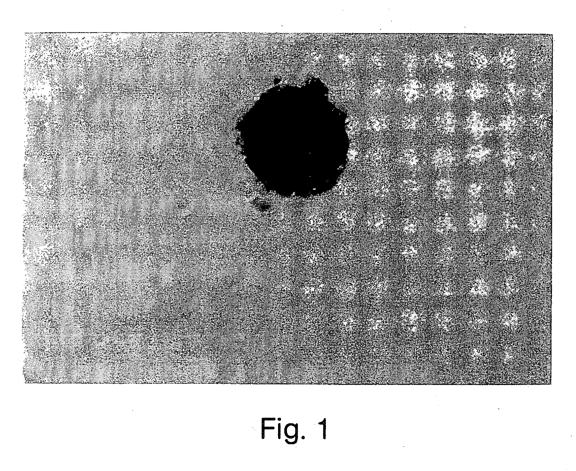
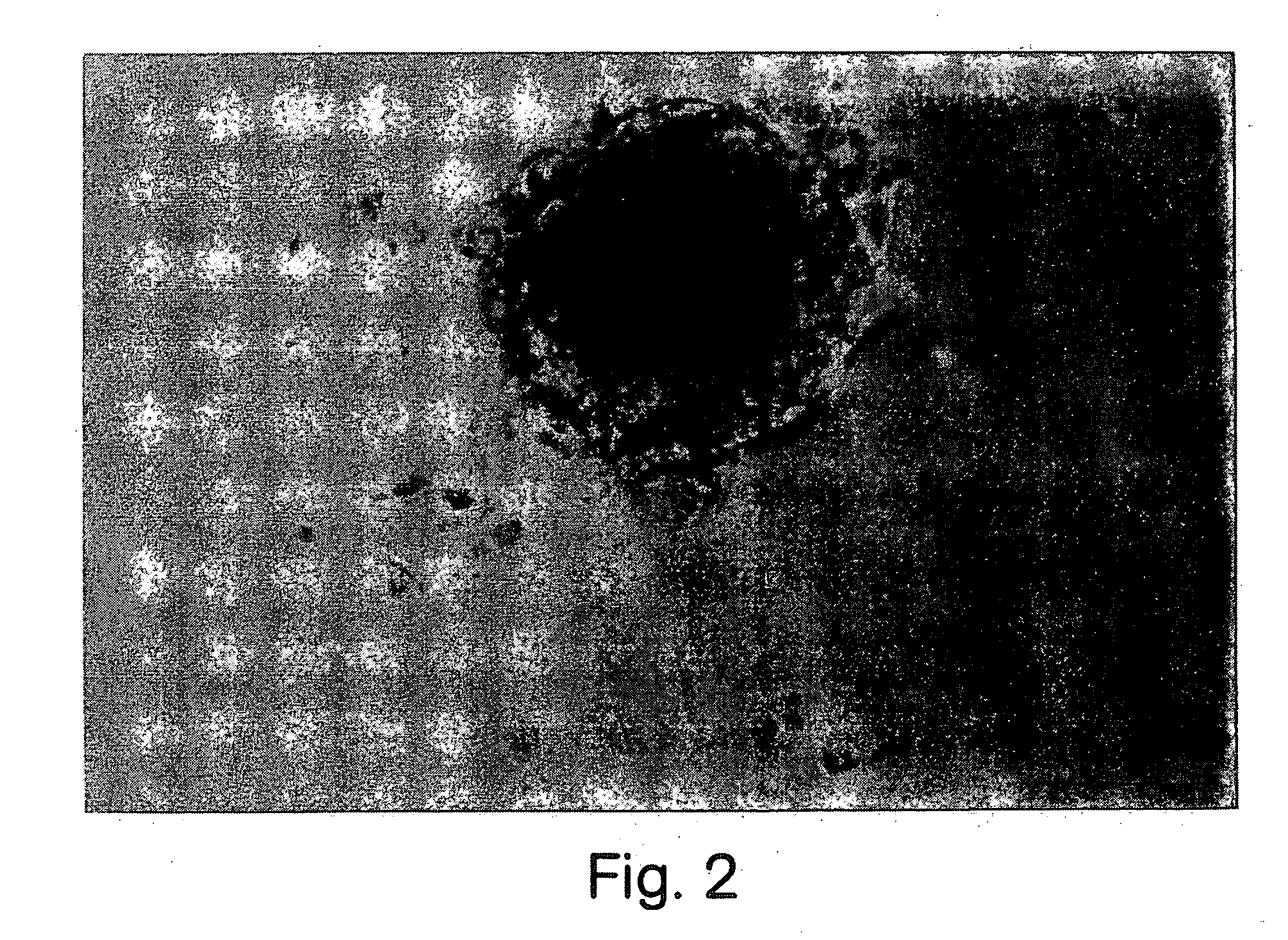
Dissection of Retina and Retinal Pigment Epithelium (RPE)

[0079] We considerably narrowed the area from which the retinal stem cell originates in the adult eye. The pigmented cells associated with the ciliary margin is the only area from which retinal spheres can be isolated in the adult animal. Dissections done on younger animals (embryonic or early post-natal) typically include the entire RPE. The cells of this invention may be used with biomaterials in a method of medical treatment of ocular dysfunction or disease. The dissection of the adult and embryonic neural retinal and RPE layers was done similarly to the method described for dissecting chick embryo retina in Opas et al., Development Biology, 161:440-454, (1993) in artificial cerebrospinal fluid (aCSF) containing 124 mM NaCl, 5 mM KCl, 1.3 mM MgCl2, 2 mM CaCl2, 26 mM NaHCO3, and 10 mM D-glucose (pH 7.4) previously aerated (15 min) with 95% O2-5% CO2 at room temperature. RPE cultures were prepared by enucleating mouse embryo...

example 2

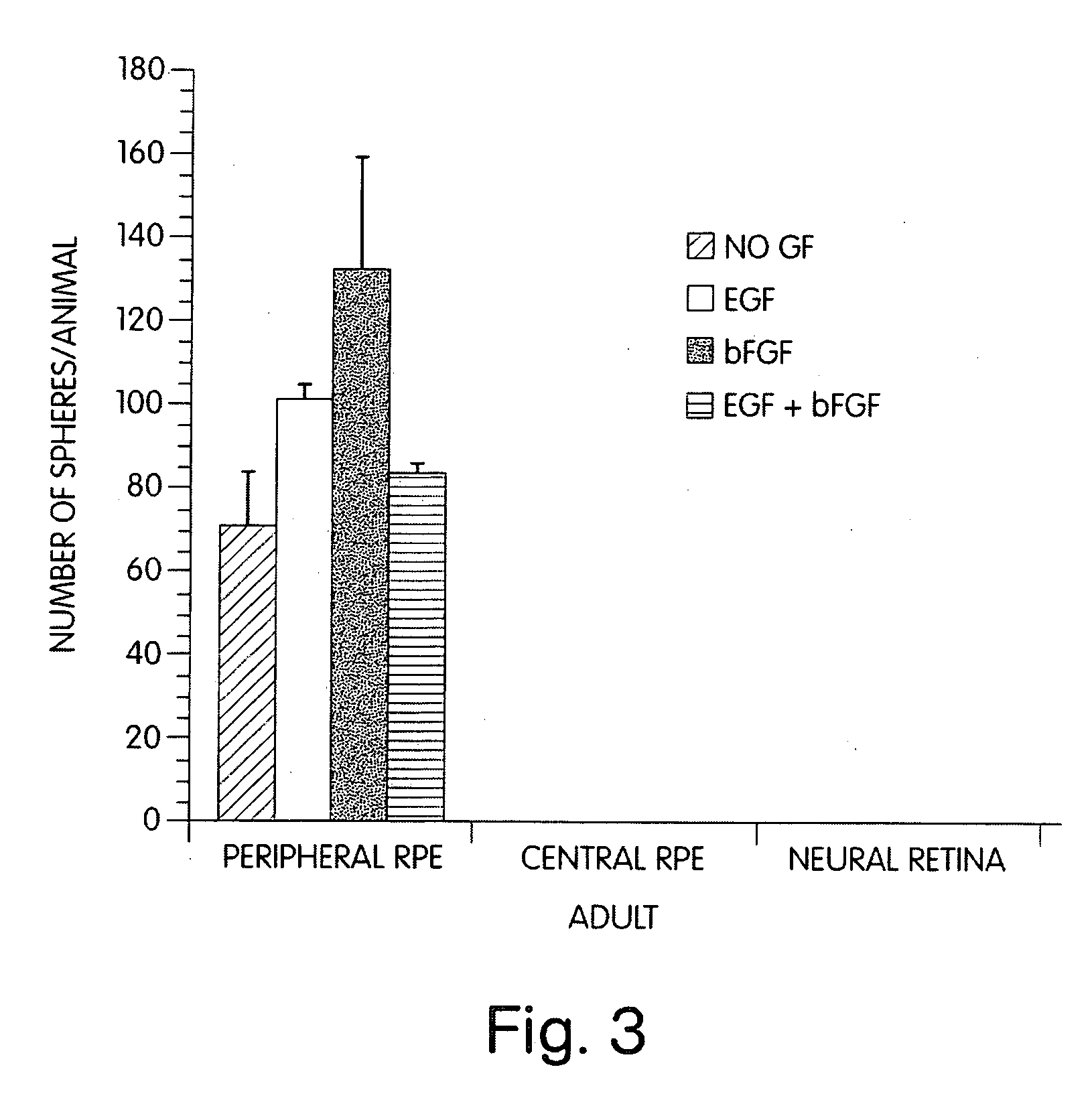
Isolation and Culturing of Retinal Stem Cells in the Presence of Growth Factors

[0080] Retinal cells were dissociated and cultured in the presence of growth factors according to the methods described in Morshead et al., Neuron, Vol. 13:1071-1082 (1994) and Reynolds et al., 1993. After dissection and enzymatic treatment, neural retina and RPE tissues were cut into 1 mm sections and transferred into serum-free culture medium (described below) containing 0.7 mg / mL trypsin inhibitor (Boehringer-Mannheim) to stop the enzymatic reaction and mechanically dissociated (trituration) with a fire-polished Pasteur pipette. The cell suspension was then centrifuged at 150×g for 5 min, the media was aspirated and the pellet was resuspended in fresh serum-free media only.

[0081] The dissociated cells were plated in noncoated 35 mm culture dishes (NUNC 96 well plates) at desired densities (determined by Trypan blue exclusion) with serum-free medium containing 20 ng / mL EGF (UBI; purified from mouse su...

example 3

ChX 10 Marker Occurs in Retinal Stem Cells

[0085] EGF, FGF2 and heparin together induced the proliferation of retinal stem cells in serum-free medium, which produced a sphere of undifferentiated precursor cells. EGF alone in serum-free medium also produced undifferentiated precursor cells. These spheres were not immunoreactive for glial fibrillary acidic protein (GFAP) (an intermediate filament protein specific for astrocytes), neuron-specific enolase (a neuron-specified enzyme), or myelin basic protein (MBP) (a cell surface protein specific to oligodendrocytes). The spheres were, however, immunoreactive for nestin (characterized by Lehndahl et al., Cell 60:585 (1990)) which is an intermediate filament protein found in undifferentiated CNS cells. The precursor cells were also immunoreactive for the Chx 10 protein marker (characterized by Liu et al., Neuron, Vol. 13:377-393 (1994); Burmeister et al. Nature Genetics 12:376-384, 1996). Chx 10 marker is a homeobox gene that is normally ...


PUM


Abstract

The invention relates to stem cells isolated from the retina of mammals and retinal cells differentiated from these stem cells. The invention also relates to a method of isolating retinal stem cells and inducing retinal stem cells to produce retinal cells. Retinal stem cells may also be induced in vivo to produce retinal cells. The invention also includes pharmaceuticals made with retinal stem cells or retinal cells which may be used to restore vision lost due to diseases, disorders or abnormal physical states of the retina. The invention includes retinal stem cell and retinal cell culture systems for toxicological assays, for isolating genes involved in retinal differentiation or for developing tumour cell lines.

Description

CROSS REFERENCE TO RELATED APPLICATION [0001] This application is a continuation of U.S. Ser. No. 09 / 333,248, filed Jun. 15, 1999, which is a continuation of U.S. Ser. No. 08 / 937,967, filed Sep. 25, 1997, abandoned, which claims priority from U.S. Provisional Application No. 60 / 026,698 filed Sep. 25, 1996, each of which is incorporated by reference herein.FIELD OF THE INVENTION [0002] The invention relates to stem cells isolated from the retina of mammals. The invention includes a method for stimulating proliferation of endogenous retinal stem cells in vivo and pharmaceutical compounds that stimulate proliferation of retinal stem cells. The invention also relates to a method for isolating retinal stem cells, uses for the stem cells and pharmaceutical compositions containing the stem cells or their progeny. The invention can be used to treat individuals having retinal diseases, disorders or abnormal physical states. The invention includes retinal stem cells and retinal cell culture s...

Claims


Application Information

Patent Timeline
IPC IPC(8): A61K35/12A61K35/44A61K38/18C12N5/071C12N5/10C12Q1/02C12Q1/18C12Q1/24
CPCA61K35/12C12N5/0621A61K35/44
Inventor KOOY, DEREK VAN DERMCINNES, RODERICKCHIASSON, BERNARDTROPEPE, VINCENZO
Owner KOOY DEREK VAN DER
Why Eureka
Try Eureka