Compositions and methods for measuring analyte concentrations

a technology of analyte concentration and composition, applied in the field of fusion proteins, can solve the problems of difficult patient compliance, inhibitors in the blood and electrodes, and easy detection of signals upon analyte binding

Inactive Publication Date: 2005-05-26
BECTON DICKINSON & CO
View PDF13 Cites 44 Cited by
  • Summary
  • Abstract
  • Description
  • Claims
  • Application Information

AI Technical Summary

Problems solved by technology

Currently, most diabetics use the “finger stick” method to monitor their blood glucose levels, and patient compliance is problematic due to pain caused by frequent (i.e., several times per day) sticks.
This method is associated with difficulties including the influence of oxygen levels, inhibitors in the blood and problems with electrodes.
In addition, detection results in consumption of the analyte that can cause difficulties when measuring low glucose concentrations.
Despite the usefulness of mutated PBPs, few of these proteins have been designed and examined, either with or without reporter groups.
Specific mutations of sites and/or a

Method used


Image

  • Compositions and methods for measuring analyte concentrations
  • Compositions and methods for measuring analyte concentrations
  • Compositions and methods for measuring analyte concentrations

Examples


example

Example 1

Preparation of Mutant GGBP and Fusion Constructs

[0071] Plasmid pTZ18R contains the MgLB gene from E. coli strain JM109. The GGBP gene was amplified from pTZ18R. The GGBP gene was ligated into the pQE70 plasmid to create a histidine-tagged protein that is wild-type in sequence, except for a lysine-to-arginine change at amino acid position 309, and the addition of a serine at amino acid position 310, before the six histidines at the C-terminus. The DsRed2 gene was amplified from (pDsRed2) and ligated to the N-terminus of the GGBP gene. A short three-alanine linker was engineered into the construct between the fluorescent protein and the histidine-tagged GGBP. Mutations of the GGBP and / or the fluorescent protein were generated in the construct by standard methods. For example, PCR was performed using primers that substitute codon(s) at or near the primary glucose contact sites. This removes the cysteine residue from the DsRed2 portion of the fusion so that when the fusion is...

example 2

Purification of Fusion Protein Comprising Mutant GGBP and DsRed2

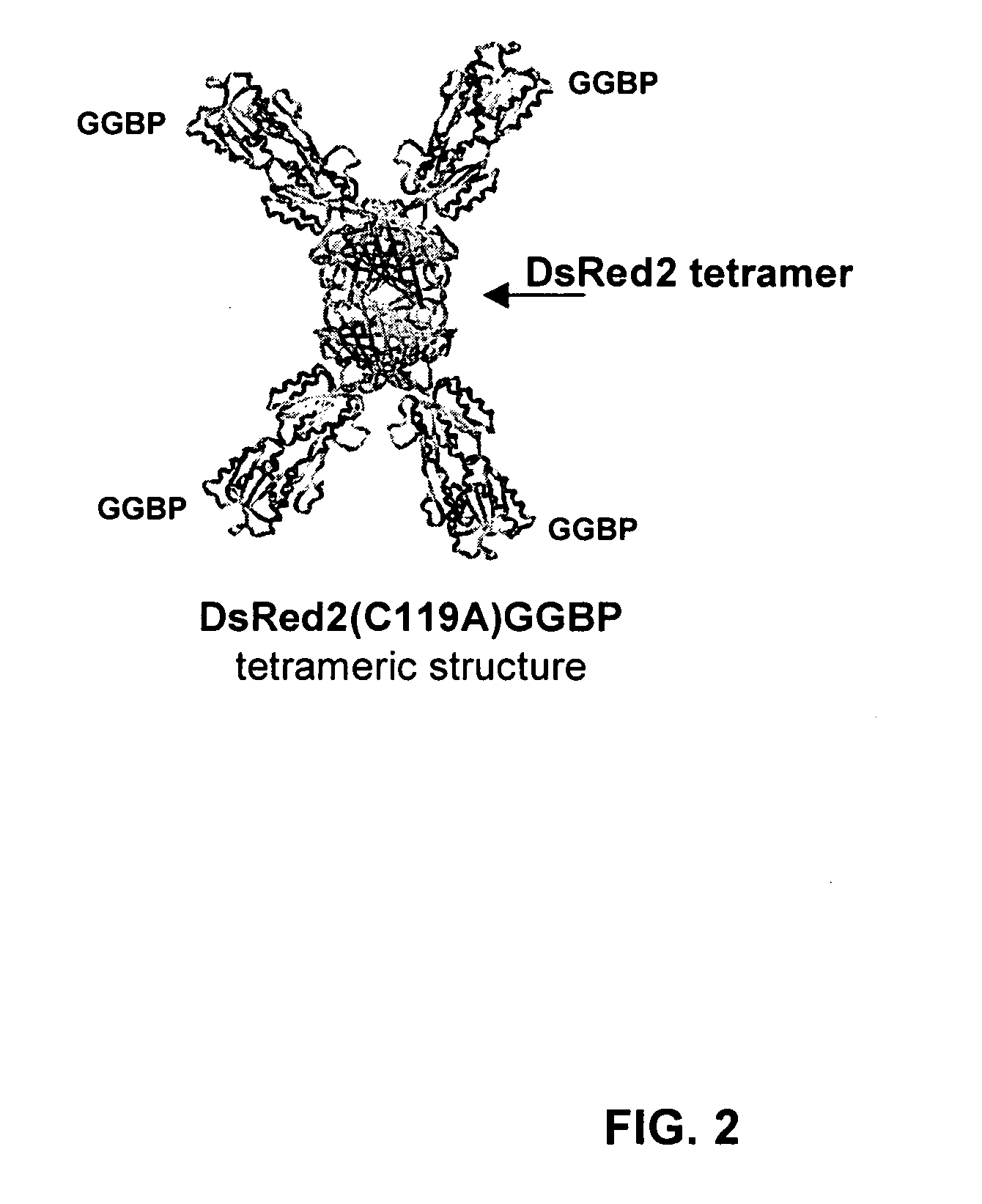
[0072] The GGBP was expressed from E. coli strain Sg13009. After E. coli induction for 72 hours, the bacteria were lysed. The lysate was cleared by centrifugation and the DsRed2(C119A)GGBP(E149C,L238C) fusion protein was purified by immobilized metal affinitive chromatography (IMAC) using Talon (cobalt-based) Resin from Clontech. The fusion protein was concentrated using a 100 kDa cutoff filter. The protein was then dialyzed at 4° C. into a solution containing 1M NaCl, 10 mM Tris-HCl, and 50 mM NaPO4 (pH 8) and stored at 20° C.

example 3

Labeling of the Fusion Protein

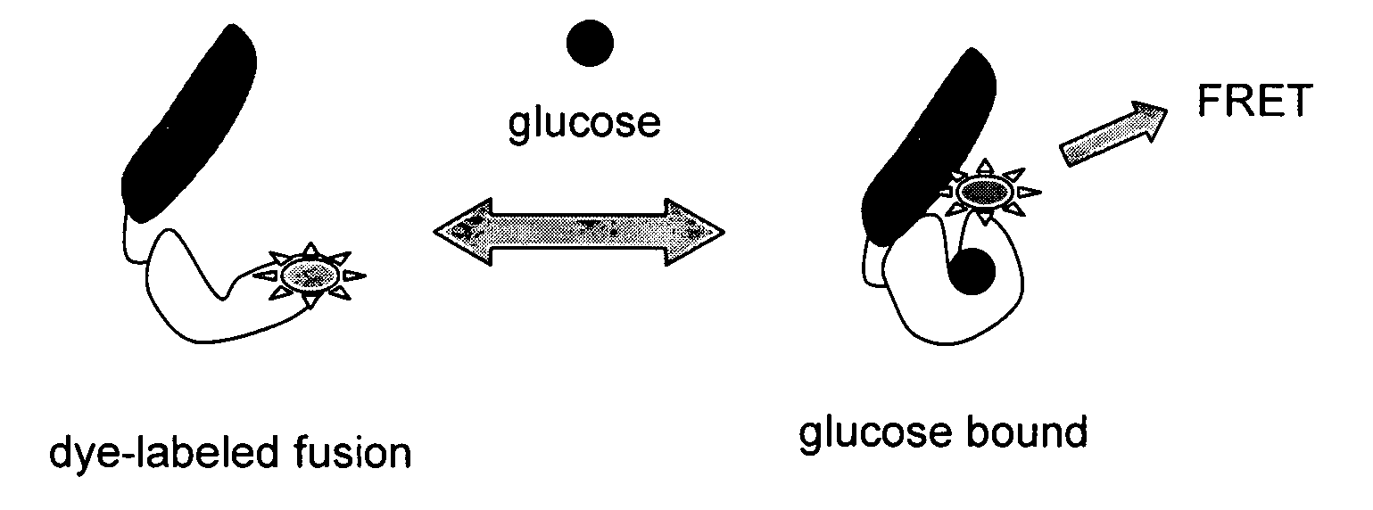
[0073] Fluorophore coupling to a thiol-reactive dye was performed by site-specifically attaching the dye through a covalent bond at a cysteine residue. The fusion was first treated with dithiothreitol, and then a 10-fold molar excess of freshly prepared fluorophore (in this case acrylodan) in dimethyl sulfoxide was subsequently added. The mixture was incubated for four hours and any unreacted dye was removed by size-exclusion column chromatography and / or dialysis. The efficiency of the coupling of the dye to the protein was determined by absorbance.


PUM


Abstract

The current invention relates to fusion proteins comprising at least one functional periplasmic binding protein, at least one labeling moiety and at least one fluorescent protein. In one embodiment, the periplasmic binding protein is a functional glucose-galactose binding protein (GGBP). The invention also relates to methods for quantifying an analyte, for example glucose, in a cell or tissue comprising administering a composition comprising a fluorescent periplasmic binding fusion protein portion to the cell or tissue, and measuring the fluorescence of the fluorescent periplasmic binding fusion protein.

Description

BACKGROUND OF THE INVENTION [0001] 1. Field of the Invention [0002] The current invention relates to fusion proteins comprising at least one functional periplasmic binding protein, at least one labeling moiety and at least one fluorescent protein. In one embodiment, the periplasmic binding protein is a functional glucose-galactose binding protein (GGBP). The invention also relates to methods for quantifying an analyte, for example, glucose, in a cell, tissue or biological fluid comprising administering a composition comprising a fluorescent periplasmic binding fusion protein portion to the cell or tissue, and measuring the fluorescence of the fluorescent periplasmic binding fusion protein. BACKGROUND OF THE INVENTION [0003] Monitoring glucose concentrations to facilitate adequate metabolic control in diabetics is a desirable goal and would enhance the lives of many individuals. Currently, most diabetics use the “finger stick” method to monitor their blood glucose levels, and patient...

Claims


Application Information

Patent Timeline
IPC IPC(8): G01N33/542G01N33/66
CPCG01N33/66G01N33/542
Inventor AMISS, TERRY J.PITNER, J. BRUCEFREITAS, TORI C.GIEL, JENNIFER L.
Owner BECTON DICKINSON & CO
Why Eureka
Try Eureka